



Retinal Detachment Surgery
Retinal detachment surgery under the care of Dr. Nishant Taneja at the Indian Eye Institute and Laser Center in Tirana. Treatment is tailored to the type and complexity of the detachment, using scleral buckling, vitrectomy, a combination of the two, or pneumatic retinopexy.

What Is Retinal Detachment?
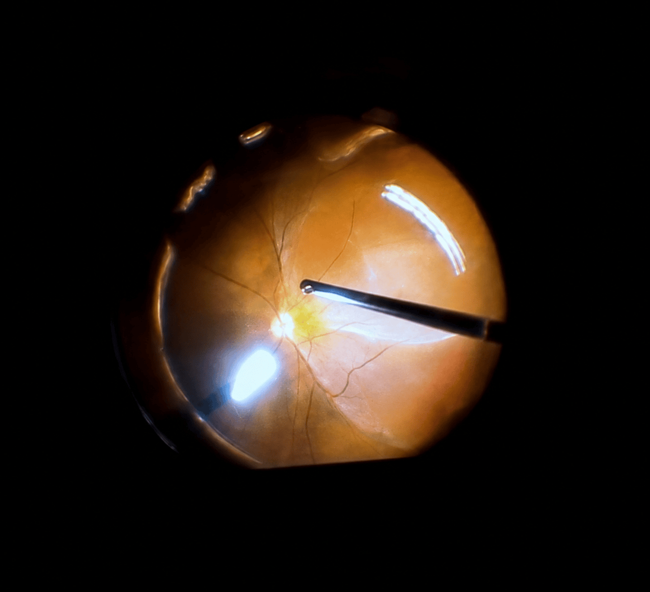
Retinal detachment occurs when the retina, the light-sensitive layer at the back of the eye, separates from the tissues that support it. This prevents it from functioning normally and can cause permanent vision loss if not treated promptly.
Most detachments are caused by a tear or hole in the retina, through which fluid passes and collects beneath it. The condition affects approximately 1 in 10,000 people each year and usually requires urgent surgical treatment.
Dr. Nishant Taneja has over 20 years of experience as a vitreoretinal surgeon and treats both straightforward and complex retinal detachments. The surgical technique is selected individually based on the characteristics of the eye and the detachment.
RETINAL DETACHMENT SURGERY AT A GLANCE
Duration:
Recovery:
Anesthesia:
Clinical experience:
PHOTO GALLERY
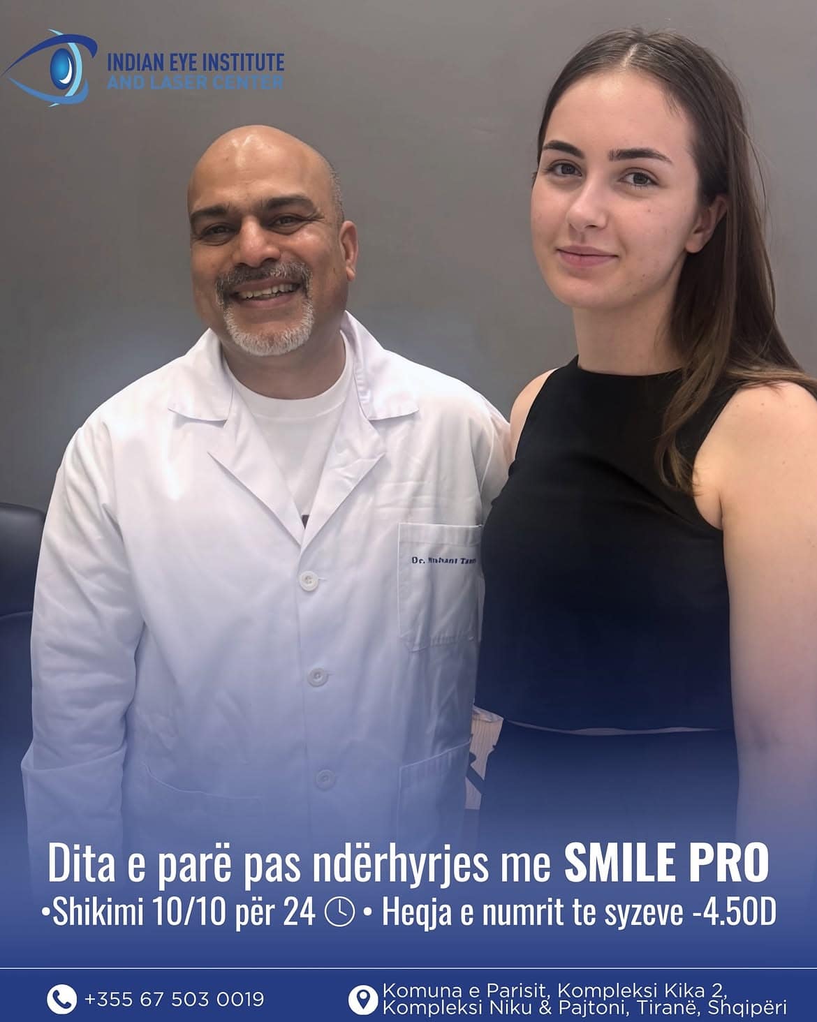
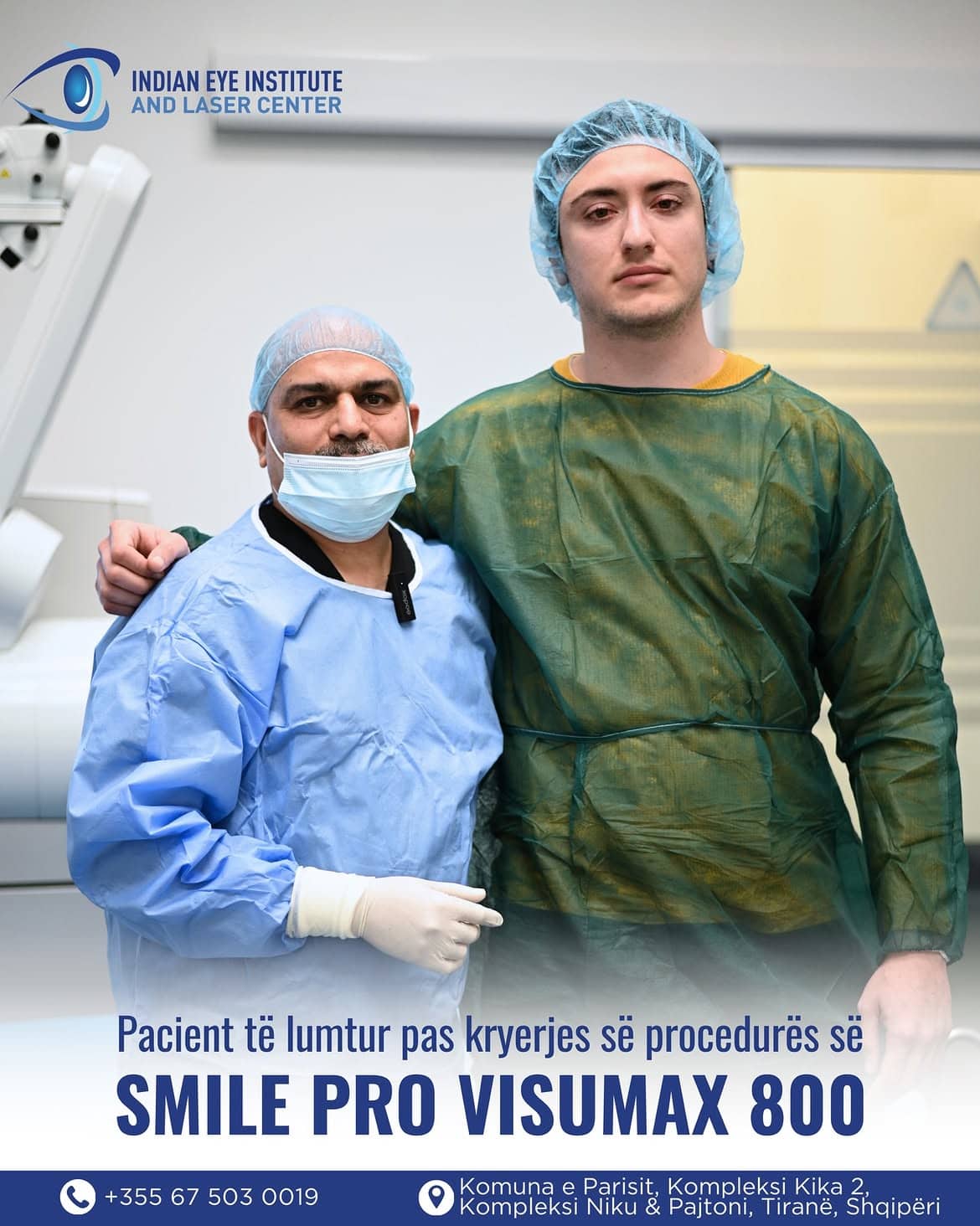
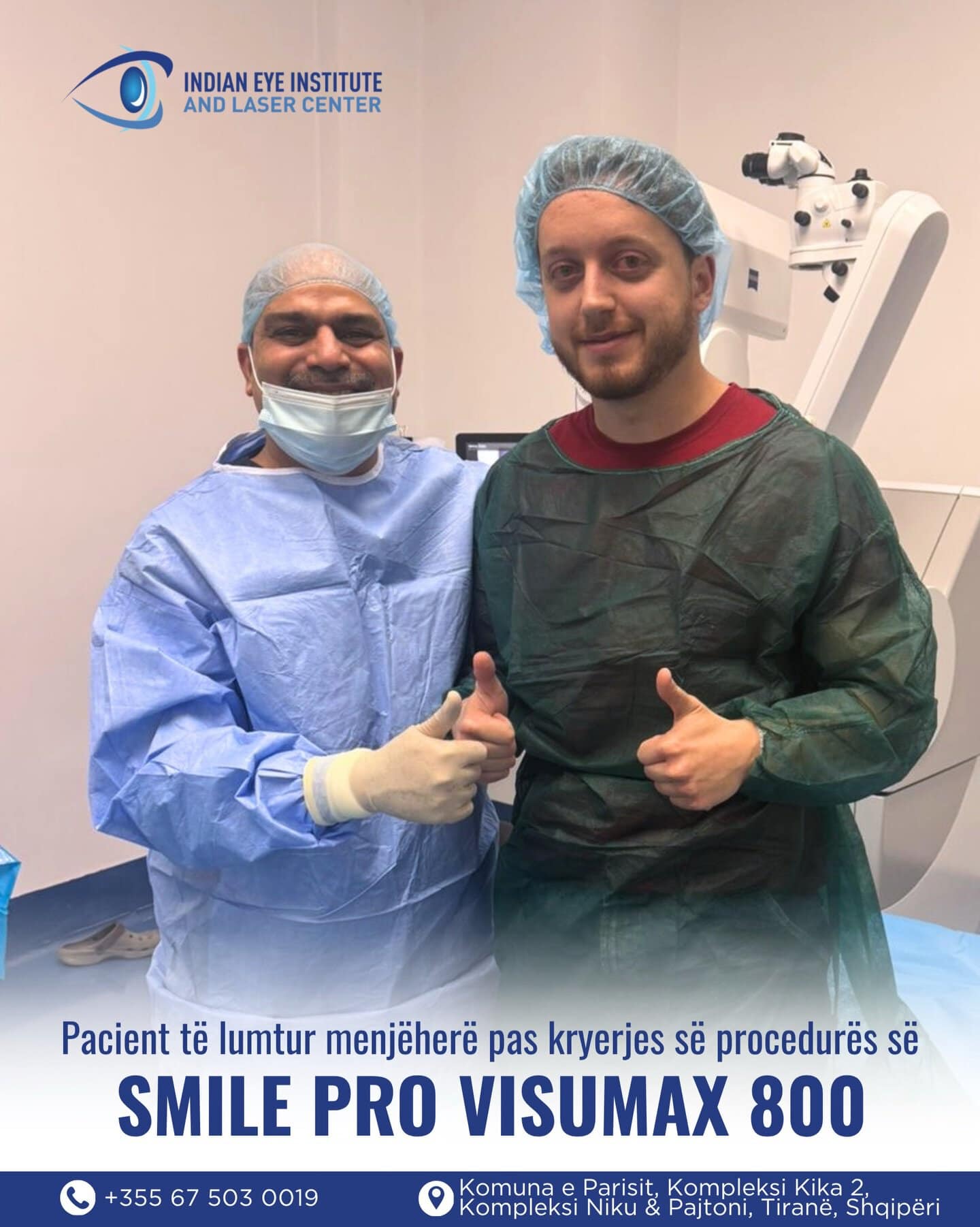
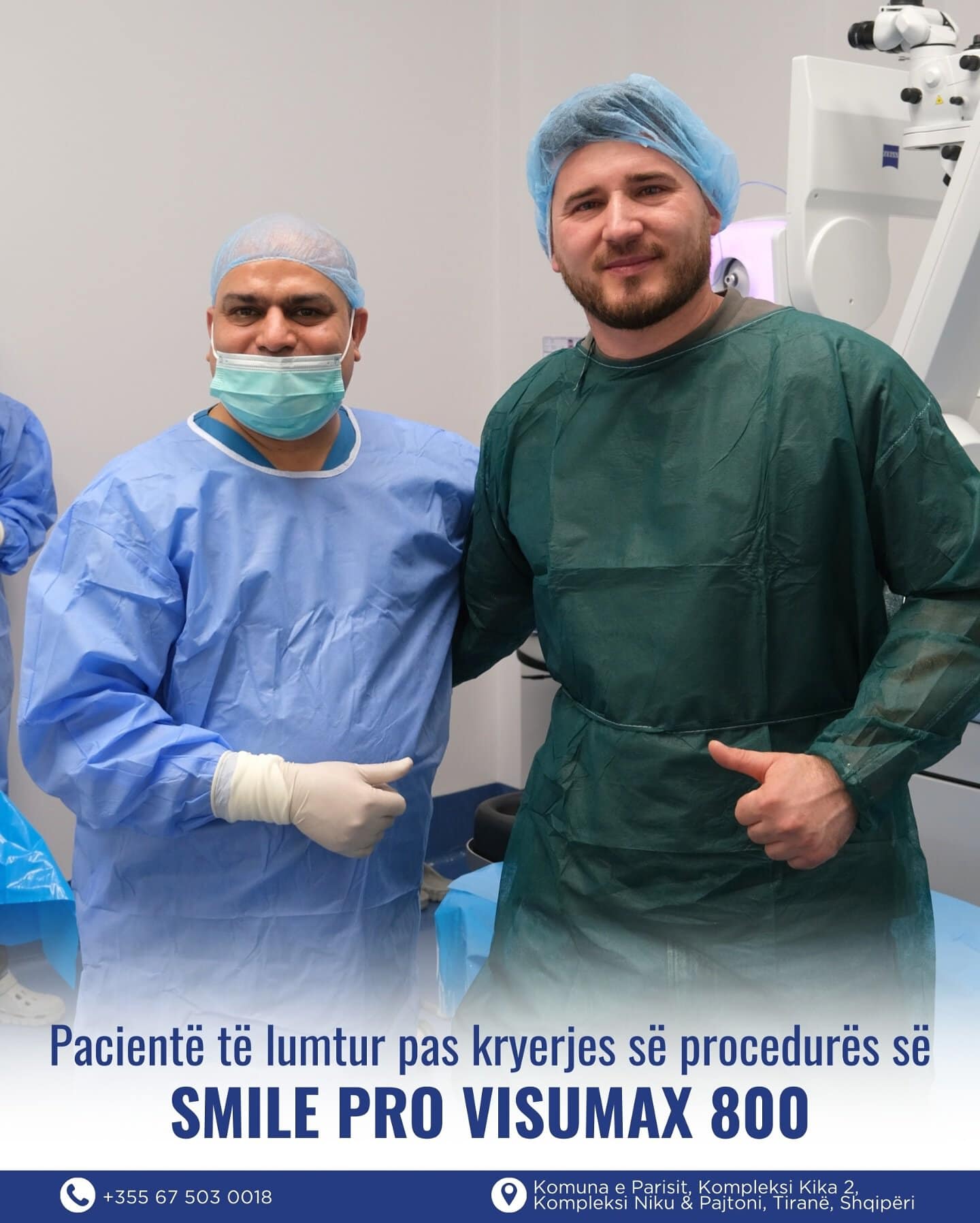
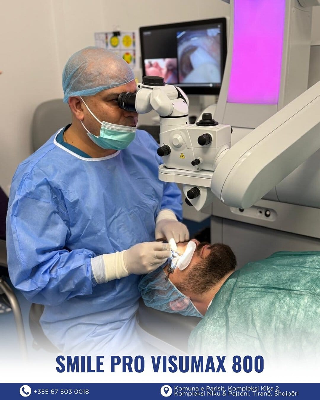
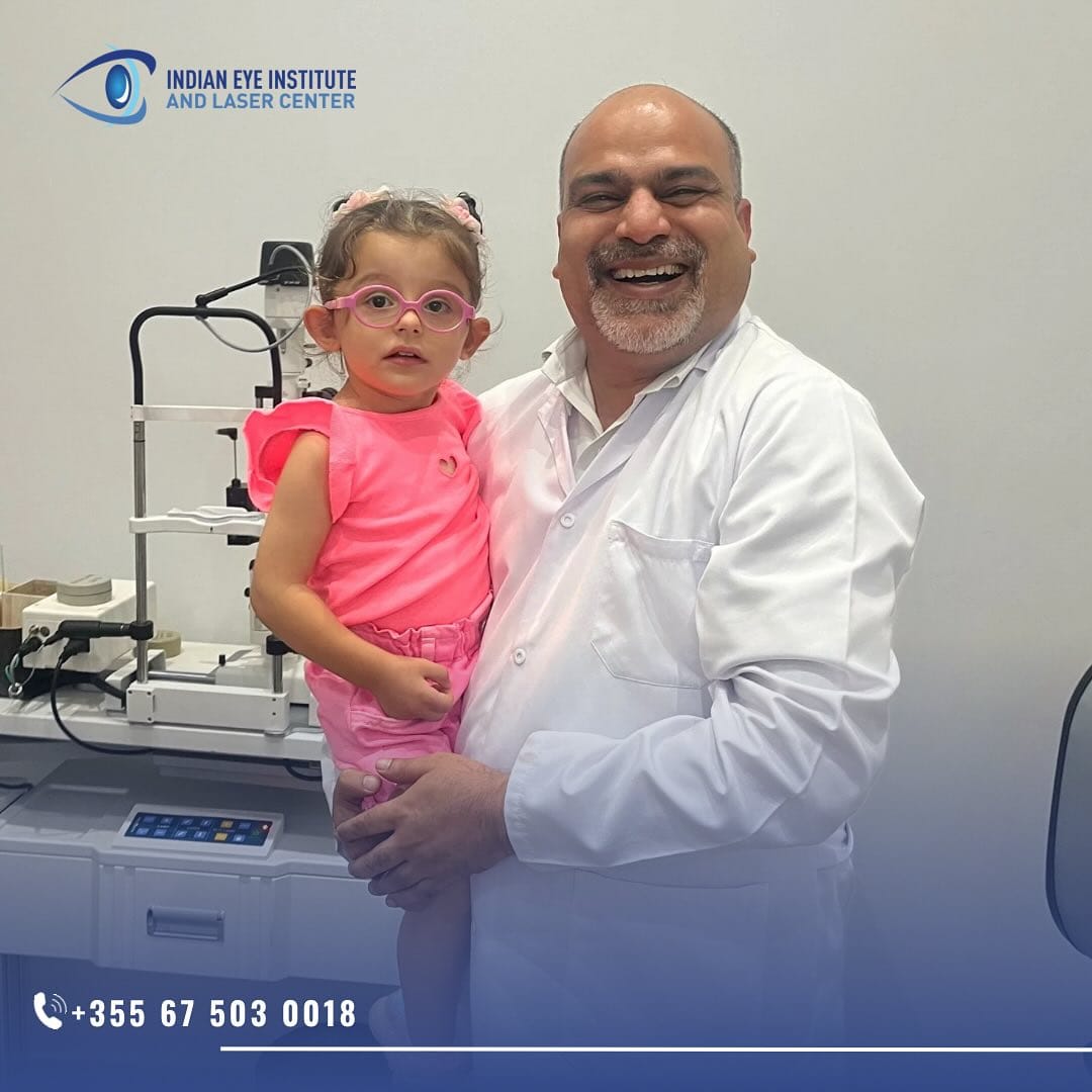
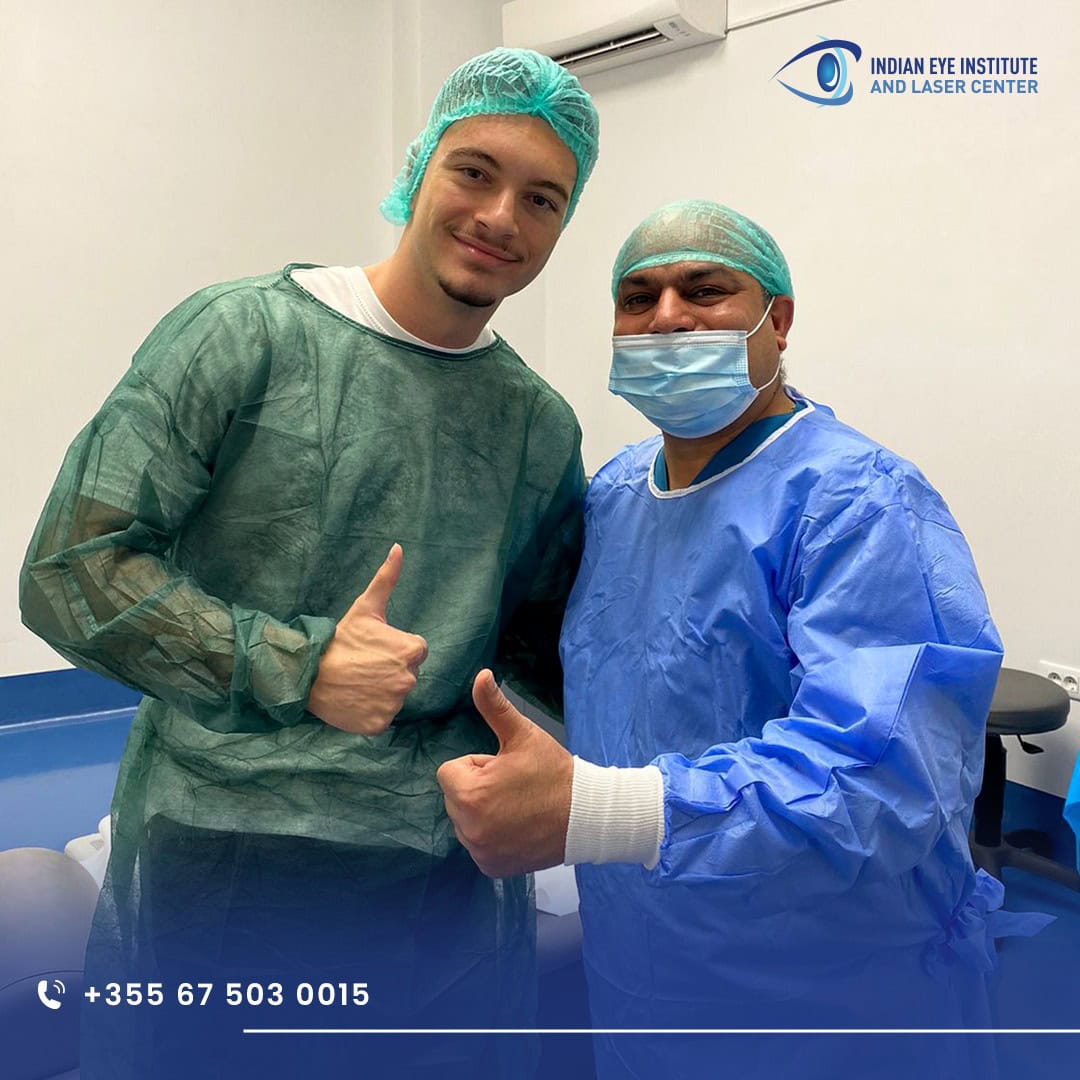
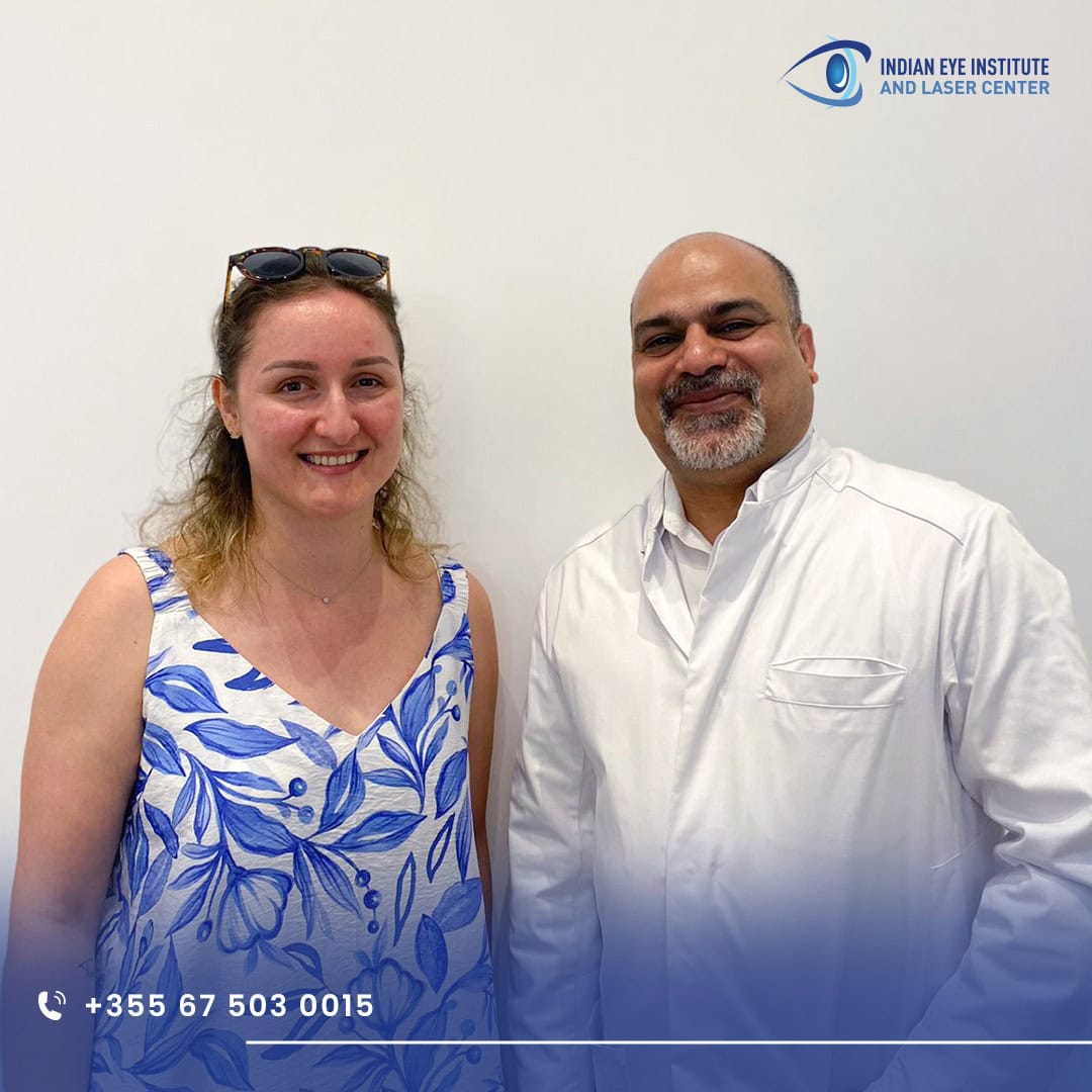
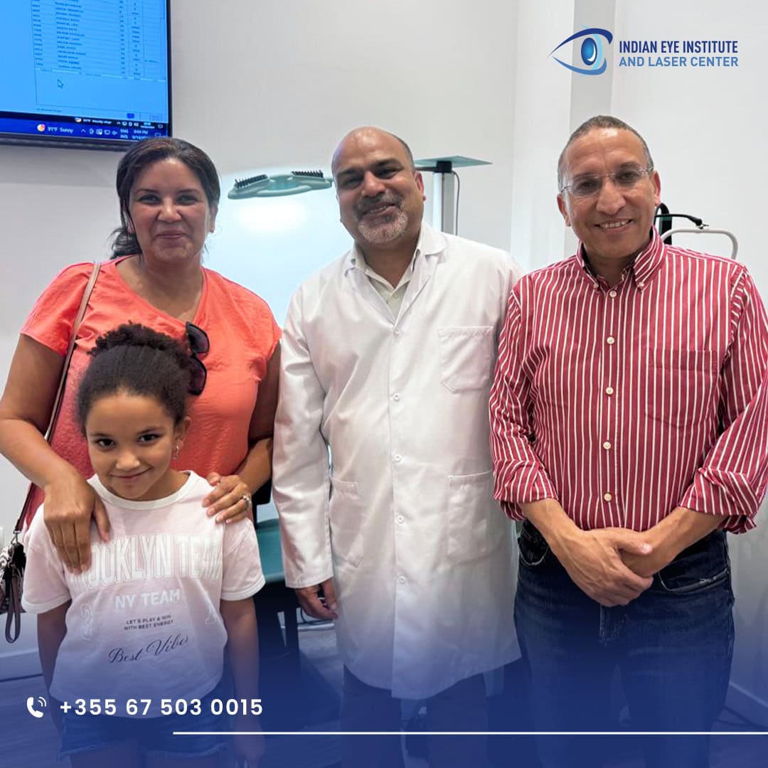
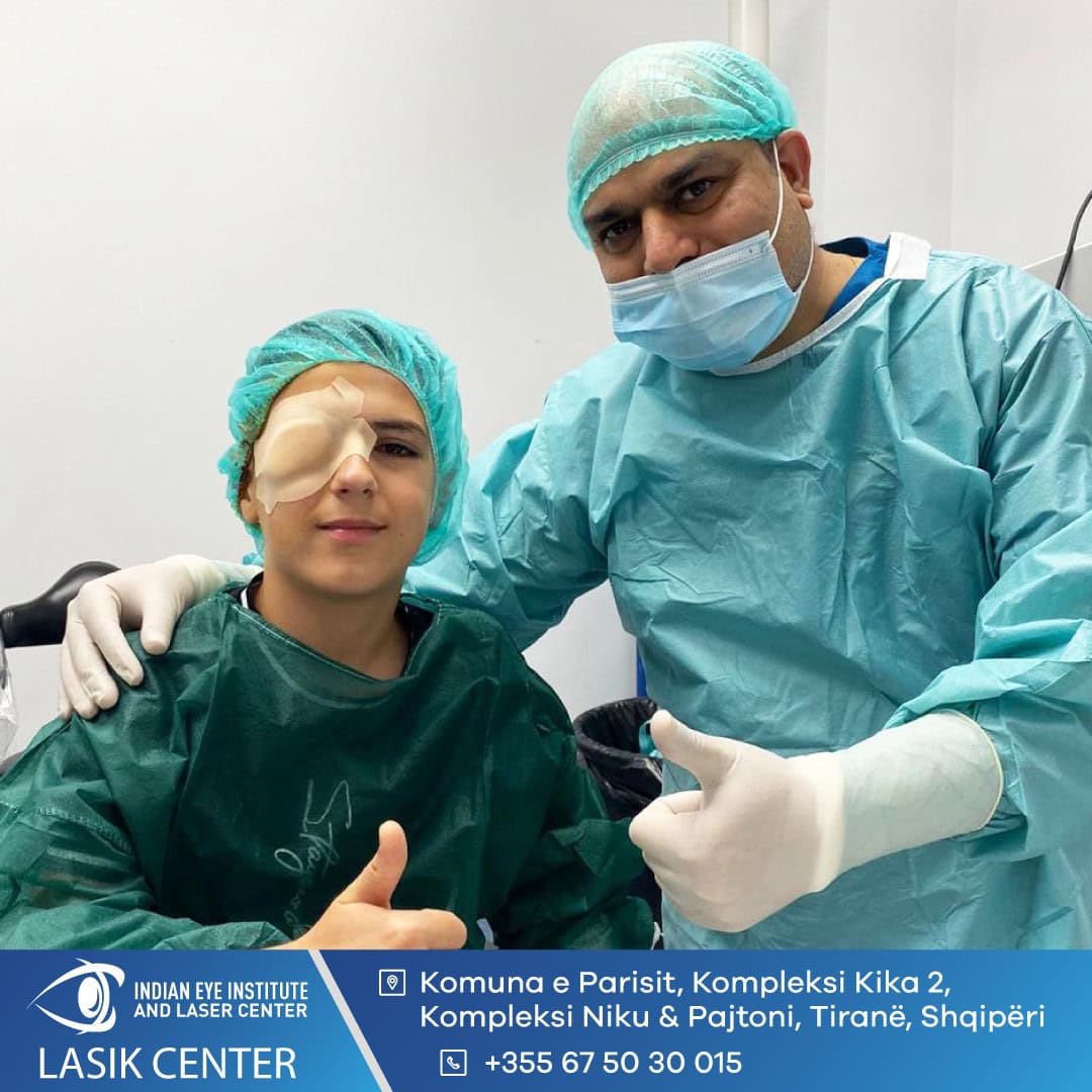
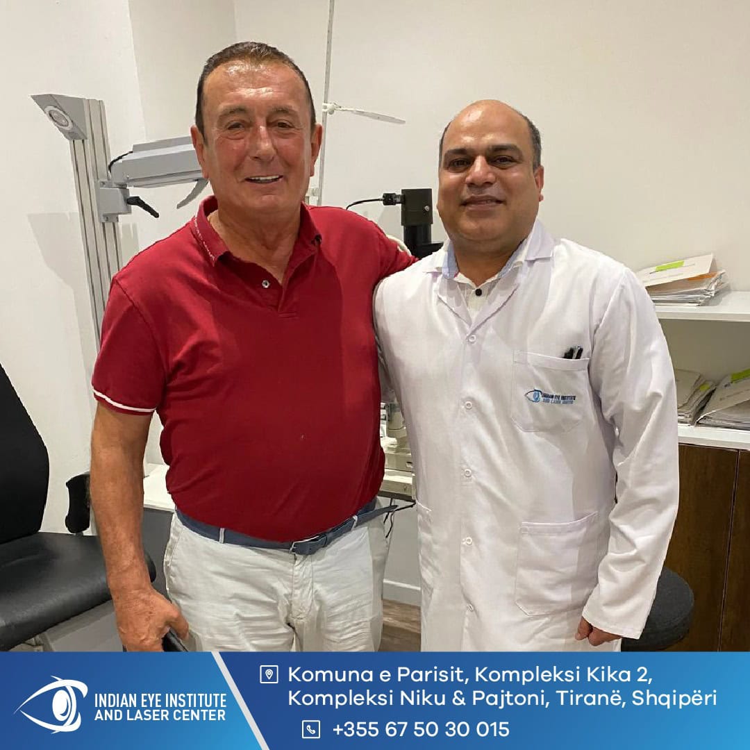
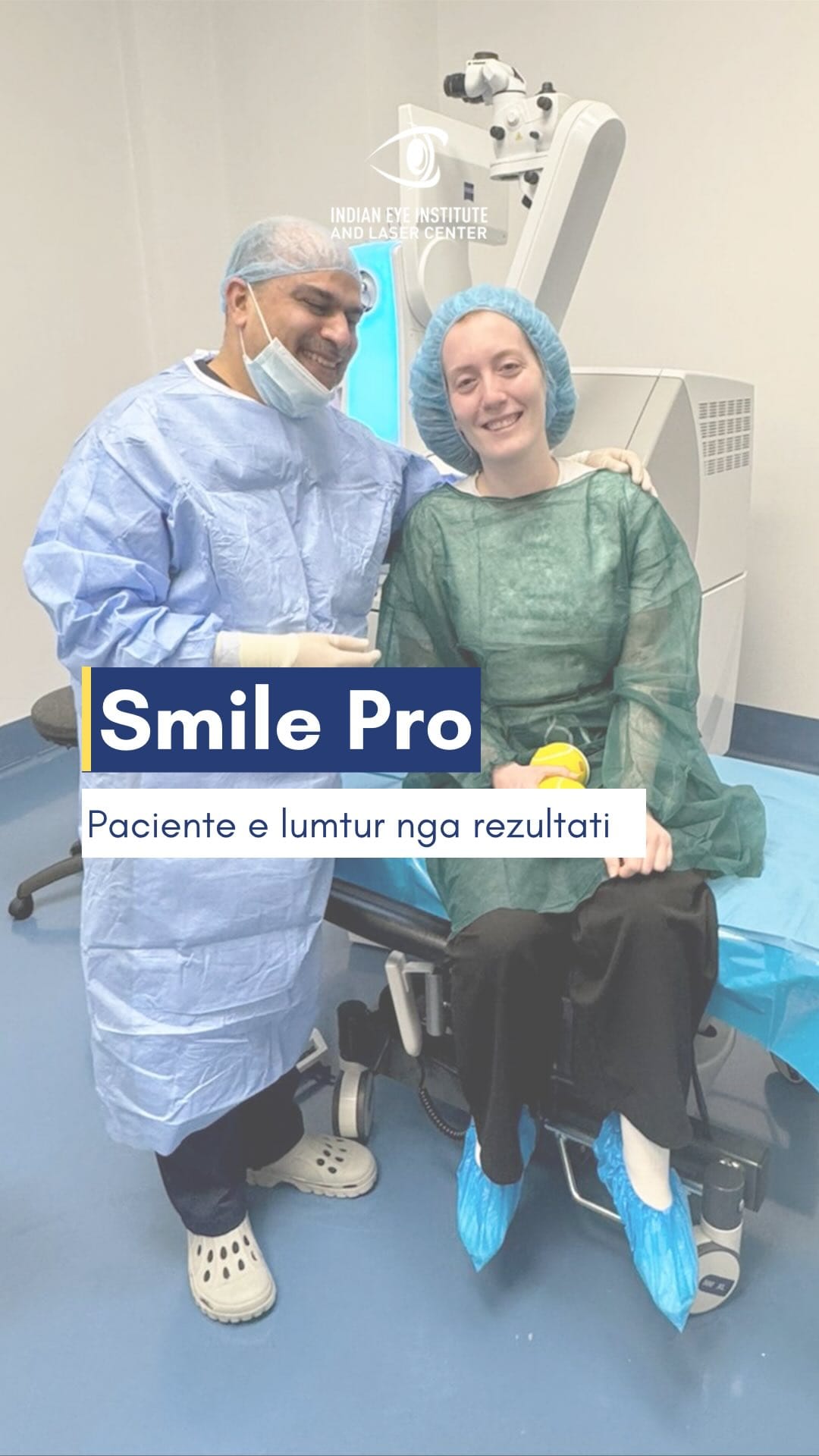
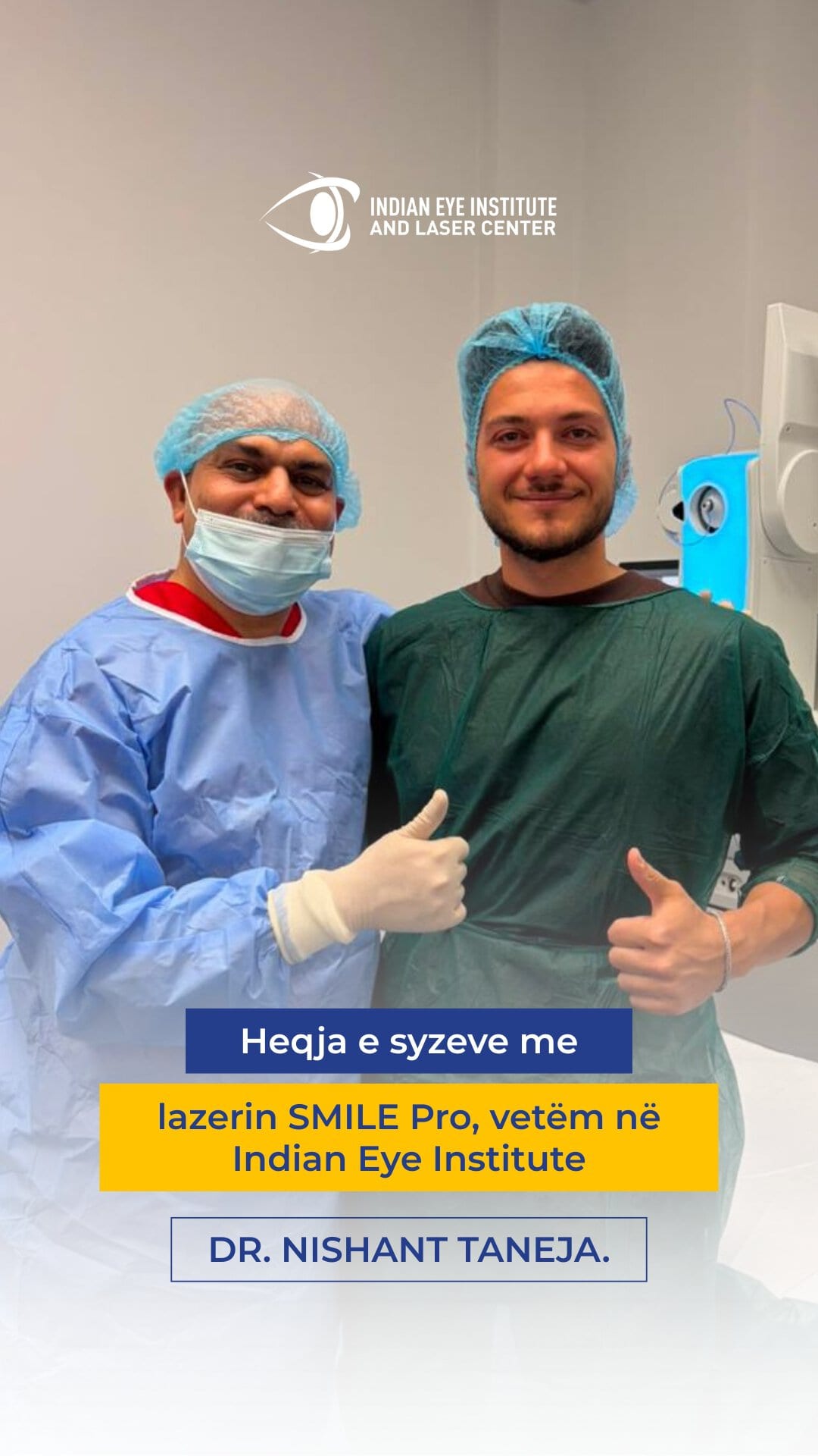
Moments with Our Patients












What Are the Symptoms of Retinal Detachment?
Symptoms may appear suddenly and are usually painless. Any such change requires an urgent examination by an ophthalmologist.
Sudden Increase in Floaters
Flashes or Sparks of Light
Dark Shadow or Curtain
Loss of Peripheral Vision
Sudden Vision Loss
No Pain
How Is Selected Treatment?
Treatment is determined by the location and number of retinal tears, the extent of the detachment, macular involvement, the presence of scar tissue, and the overall condition of the eye.
Location of the Tears
The position of the tears influences the choice between scleral buckling, vitrectomy, and pneumatic retinopexy.
Macular Involvement
If the detachment has reached the macula, the final visual outcome may depend on the duration and extent of the damage.
Complexity of the Detachment
Detachments involving multiple tears, scar tissue, or previous surgery may require a combination of scleral buckling and vitrectomy.
With extensive experience treating both simple and complex retinal detachments, Dr. Taneja determines the most appropriate technique for each patient. He also trains the next generation of vitreoretinal surgeons in these procedures.
Can prevented Retinal Detachment?
Not all cases can be prevented. However, regular eye examinations and early treatment of retinal tears can reduce the risk of a more extensive detachment.
Have regular eye examinations: Routine examinations are especially important for people with high myopia, previous eye surgery, or a family history of retinal detachment.
Treat retinal tears early: Tears or holes detected during an examination can be treated with laser therapy or cryotherapy to limit fluid from passing beneath the retina.
Protect your eyes from injury: Wear protective eyewear during work or sports activities that could result in impact or injury to the eye.
Respond promptly to symptoms: The sudden appearance of floaters, flashes, or a dark curtain requires an urgent examination by an ophthalmologist.
Benefits of Retinal Detachment Surgery
Surgery aims to reattach the retina and prevent further vision loss. The outcome depends on the type of detachment, whether the macula is involved, and how promptly treatment is performed.
Retinal Reattachment
Preservation of Vision
Treatment of the Causative Tears
Personalized Surgical Technique
High Anatomical Success Rate
Surgical Techniques for Retinal Reattachment
Scleral Buckling
A silicone band or sponge is placed on the white part of the eye to bring the eye wall closer to the detached retina. The material is covered by the conjunctiva and is not visible externally.
- Placement of a silicone band or sponge
- Treatment of retinal tears with cryotherapy
- Supporting the retina from the outside
- Leaving the buckle in place permanently, when appropriate
Pars Plana Vitrectomy
Vitrectomy is performed through several micro-incisions and involves removing the vitreous gel, treating the retinal tears, and reattaching the retina.
- Removal of the vitreous gel
- Removal of fluid from beneath the retina
- Treatment of retinal tears with laser or cryotherapy
- Placement of gas or silicone oil
Combined Treatment or Pneumatic Retinopexy
For more complex detachments, scleral buckling and vitrectomy may be combined. In selected cases, a gas bubble may be used to hold the retina in place.
- Combining scleral buckling with vitrectomy
- Injection of a gas bubble into the eye
- Sealing retinal tears with laser or cryotherapy
- Maintaining a specific position after the procedure
What to Expect
01. Retinal Examination
During the examination, visual acuity is assessed, intraocular pressure is measured, and the eye is examined with a slit lamp. After the pupils are dilated, the entire retina is carefully examined to identify tears, holes, and detached areas.
Depending on the condition, retinal photography, macular OCT, or ocular ultrasound may be performed. Ultrasound is particularly useful when a cataract, bleeding, or clouding of the eye’s structures prevents a direct view of the retina.
02. Procedure
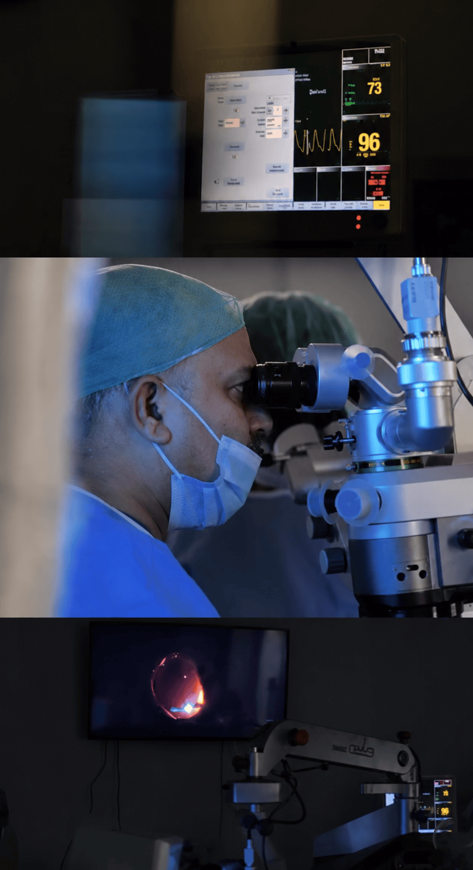
The procedure is usually performed under local anesthesia with sedation. The patient remains relaxed and should not feel pain during surgery.
Depending on the characteristics of the detachment, Dr. Taneja uses scleral buckling, pars plana vitrectomy, a combination of the two, or pneumatic retinopexy. Retinal tears are sealed with laser or cryotherapy, and the retina is supported with gas, silicone oil, or a scleral buckle.
Once the procedure is complete, the eye is covered and the patient is monitored. Instructions regarding positioning, medications, and follow-up appointments are provided before the patient leaves the clinic.
03. Recovery and Follow-Up
After surgery, blurred vision, redness, tearing, or a scratchy sensation may occur. Improvement takes place gradually over several weeks, and the final outcome varies from patient to patient.
If gas or silicone oil has been used, the patient may be asked to keep their head in a specific position. Depending on the case, positioning may be required for up to 20 days and must be followed exactly as instructed by the surgeon.
While a gas bubble remains in the eye, the patient must not travel by air, ascend to high altitudes, or receive nitrous oxide anesthesia until the gas has been fully absorbed. If silicone oil has been used, a second procedure may be required to remove it.
Severe pain, worsening redness, discharge, or a sudden decrease in vision requires immediate contact with the clinic.


Indian Eye Clinic Tirana: 15+ Years of Trust


Retina | Cataract | Refractive Surgery


The right care for a lifetime with clear vision
Every successful treatment begins with an accurate diagnosis and a personalized care plan. Discover the real stories of patients who trust Indian Eye Institute and Laser Center.


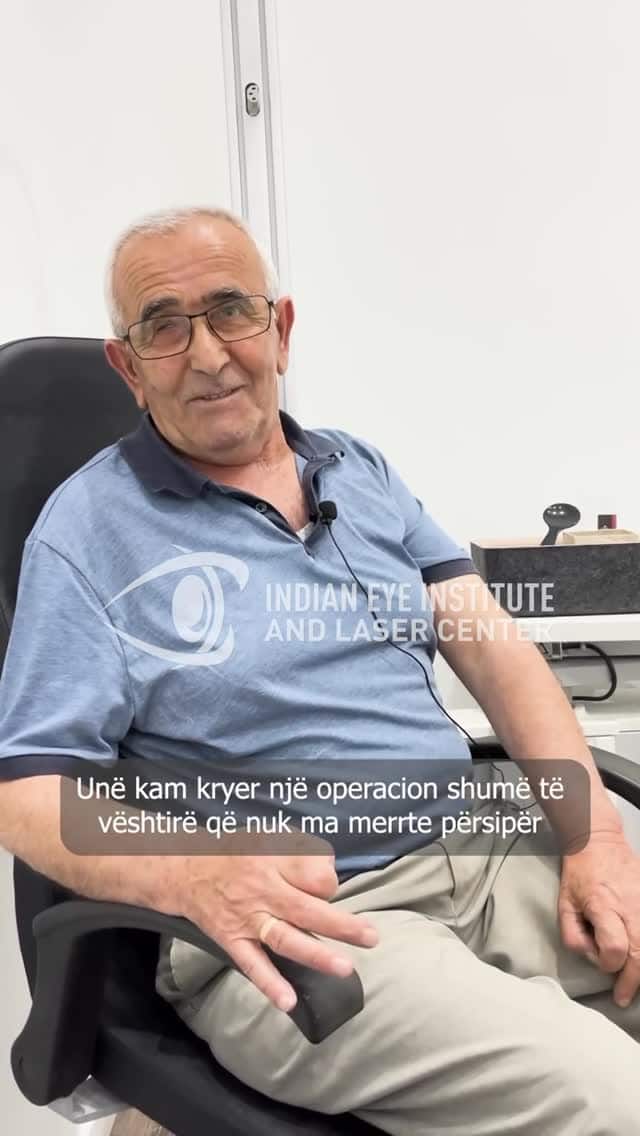


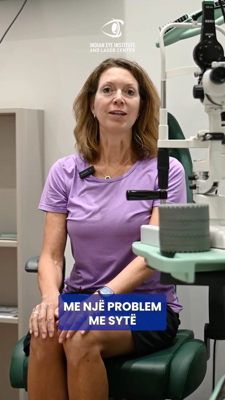

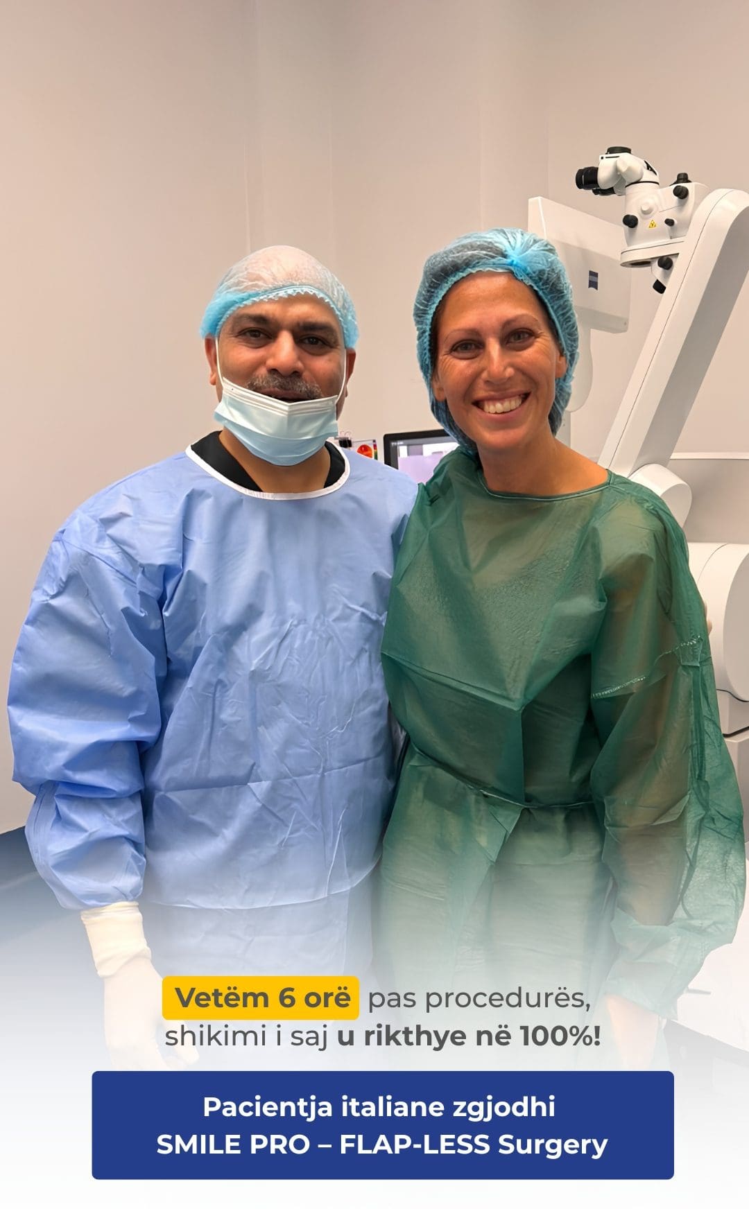

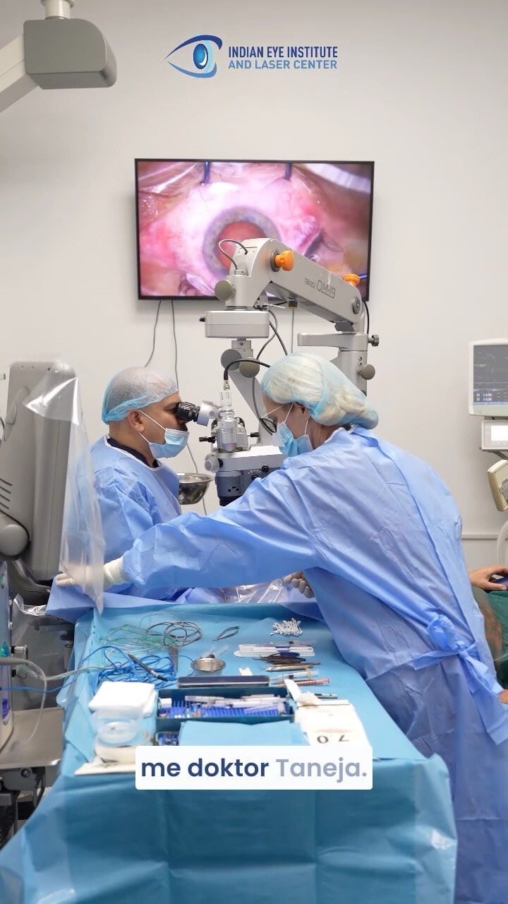
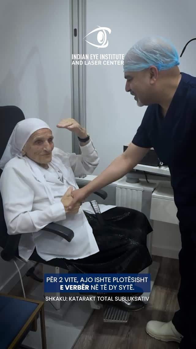



I have advanced glaucoma in both eyes and have undergone glaucoma and cataract surgery with Dr. Taneja. I have been receiving regular follow-up care from Dr. Taneja for the past 11 years.
I am very happy and satisfied with my experiences at Dr. Taneja’s clinic. The staff are helpful, knowledgeable, competent, and very pleasant. Dr. Taneja has worked diligently to preserve my vision, and I am extremely pleased with the care I have received. He deserves recognition for how good he is at what he does. I would recommend him 100% to anyone with serious eye problems.
I have suffered from a long-term problem with floaters in both eyes. Dr. Nishant Taneja is truly an expert in his field who allows patients to consider nonsurgical options as the first course of treatment. He is professional and courteous, treats patients with respect, and communicates clearly in terms that ordinary people can understand. I believe he is honest and straightforward, and he is taking excellent care of me.
I have been a patient of Dr. Nishant Taneja since 2010, when I was diagnosed with wet macular degeneration. He has treated me with Avastin, Eylea, and now brolucizumab, and I can still read large print.
I owe my sight to his treatments. He has taken excellent care of my eyes. The staff are absolutely outstanding in their courtesy and professionalism. I am 93 years old and can still read clearly. Thank you!
When I first came here, I thought I would go blind in my left eye. I was diagnosed with a vitreous hemorrhage caused by detachment of the vitreous gel. After the laser treatment, my vision returned to what it had been before.
I have received excellent medical care here. The entire staff have been very kind and have made everything much easier for me. I am very happy.
Many thanks to the entire staff. The whole process of my cataract surgery and the care I received were the best I have ever experienced at any medical practice. The staff and technology were first-rate. Dr. Taneja is simply phenomenal. Thank you again.
I underwent surgery for a retinal detachment last week. I experienced no pain during or after the operation. I slept well the following night without needing any pain medication. If anyone has questions about retinal surgery, please feel free to call me.
I am impressed by the high standard of patient care I received from Dr. Taneja at the Indian Eye Institute. I hold him in the highest regard.
I have had diabetes for over 30 years. Over the past 5 years, my vision gradually deteriorated. I had been receiving treatment from my ophthalmologist for the previous 4 years. The treatment involved injections, but the situation seemed to be getting worse. I could barely see well enough to drive, could not read anything on my phone, and began having difficulty recognizing people from a distance. I therefore decided to visit Dr. Nishant Taneja on the recommendation of my eye doctor. He was prompt, efficient, and professional. Within minutes, he had mapped out a treatment plan for both of my eyes and advised me to follow it. I was truly impressed by his energy, optimism, and confidence.
After several injections and laser treatments, he said that I needed a vitrectomy in my left eye. It went perfectly, without any complications. My vision in that eye was restored to 20/20. I felt as though I were opening my eyes for the first time.
After I recovered from that procedure, Dr. Taneja said that my right eye also needed surgery. So, without hesitation, I had that eye treated as well. I now have 20/20 vision in both eyes, and everything is wonderful.
I can now see my two young grandchildren clearly. This has improved my quality of life tremendously. Without a doubt, I am probably the happiest patient Dr. Taneja has ever had!
I am delighted to express my appreciation for Dr. Taneja and his exceptional staff.
If you are reading this review while deciding on a retina specialist, you have definitely come to the right place! An excellent doctor and staff. I am more than completely satisfied!
I believe Dr. Taneja is an exceptionally talented surgeon.
I began wearing glasses and contact lenses at the age of 12. I relied on them to see and read. During the spring, pollen irritated my eyes, and I could no longer wear my contact lenses.
After the procedures performed by Dr. Taneja, I went from being unable to see to having clear vision without contact lenses or glasses.
I would recommend Dr. Nishant Taneja for any procedure he performs. He is highly meticulous, compassionate, and understanding.
He worked a miracle for me. I thank God for his talent.
Patient Education Videos
View All
The SMILE Pro Procedure
Chemical Eye Injuries
Diabetic Retinopathy
Signs That Should Not Be Ignored
Examination and Treatment
Download the Guides Free for Patients
Frequently Asked Questions
Yes. Retinal detachment can progress rapidly and cause permanent vision loss. The onset of flashes, numerous floaters, or a dark curtain in your field of vision requires immediate examination.
The procedure is usually performed under local anesthesia with sedation, so the patient should not feel pain during surgery. Mild discomfort, a scratchy sensation, or redness may occur after the procedure.
Anatomical reattachment of the retina does not guarantee full restoration of vision. The outcome depends on the extent and duration of the detachment, whether the macula is involved, and the condition of the retina before surgery.
Positioning helps the gas bubble or silicone oil apply pressure to the treated area and keep the retina in place while it heals. The required position and duration are determined individually.
Take the First Step Toward Better Vision.
Fill out the form below and our team will contact you
to schedule a consultation and answer your questions.