



Keratoconus
Diagnosis and treatment of keratoconus under the care of Dr. Nishant Taneja at the Indian Eye Institute and Laser Center in Tirana. Using corneal topography, specialized lenses, corneal collagen cross-linking, and other treatments, care is tailored to the stage and progression of the condition.


What Is Keratoconus?
Keratoconus is a progressive disorder in which the cornea thins, weakens, and changes from its normal round shape to a cone-like shape. This process is known as corneal ectasia.
The cornea refracts and focuses light onto the retina. When its shape becomes irregular, vision may become blurred or distorted, making it more difficult to see objects clearly at both near and far distances.
Keratoconus usually develops during adolescence or early adulthood and may progress more rapidly in younger patients. The exact cause is unknown, but it is thought to involve genetic and environmental factors. Persistent, vigorous eye rubbing may contribute to its progression.
Early diagnosis and monitoring are important, as timely treatment can help slow or halt further weakening of the cornea.
CORNEAL COLLAGEN CROSS-LINKING AT A GLANCE
Duration:
Recovery:
Anesthesia:
Clinical experience:
PHOTO GALLERY
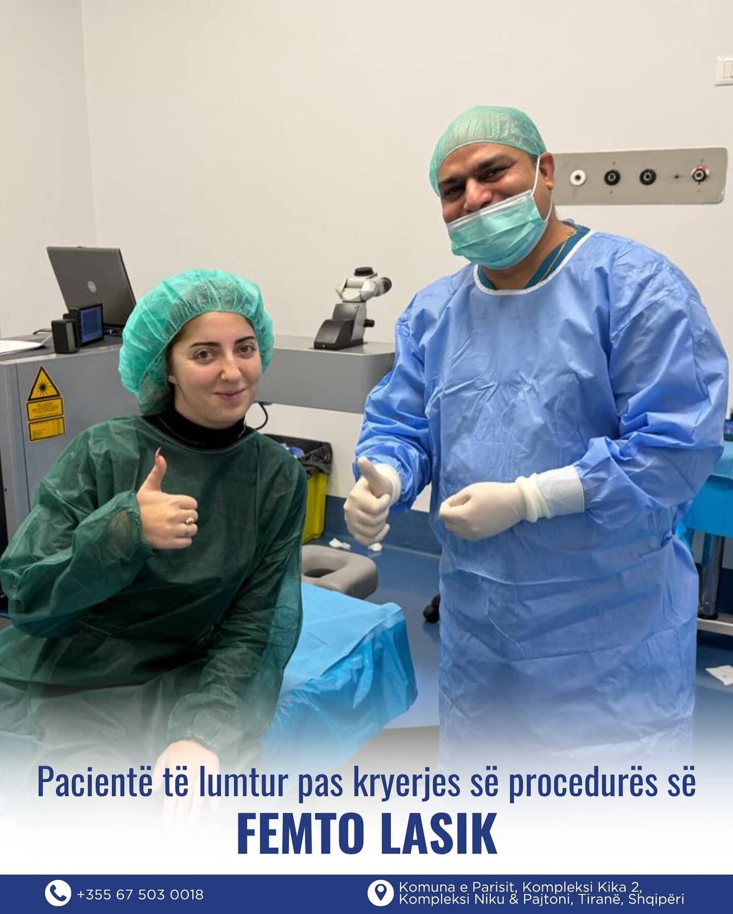
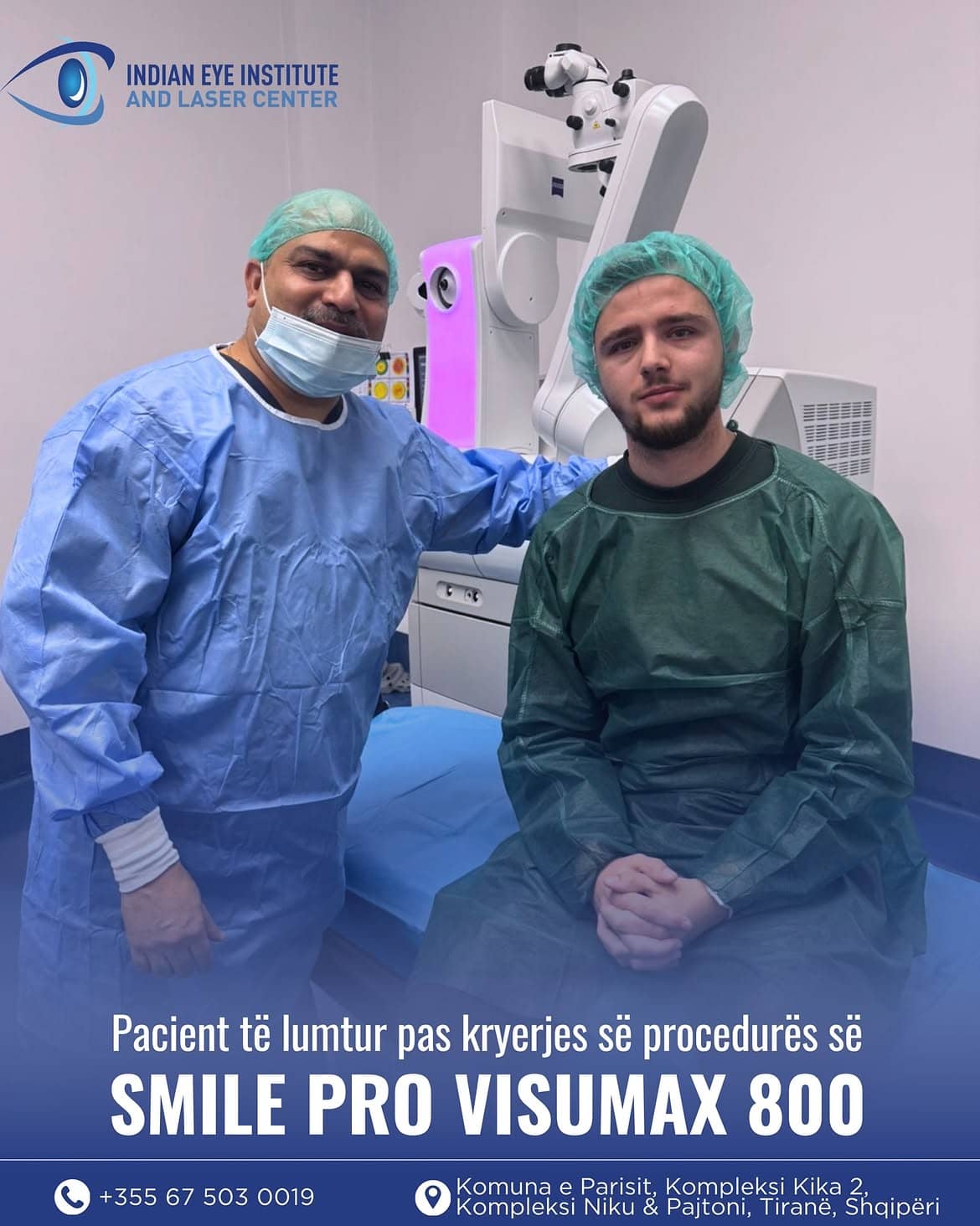
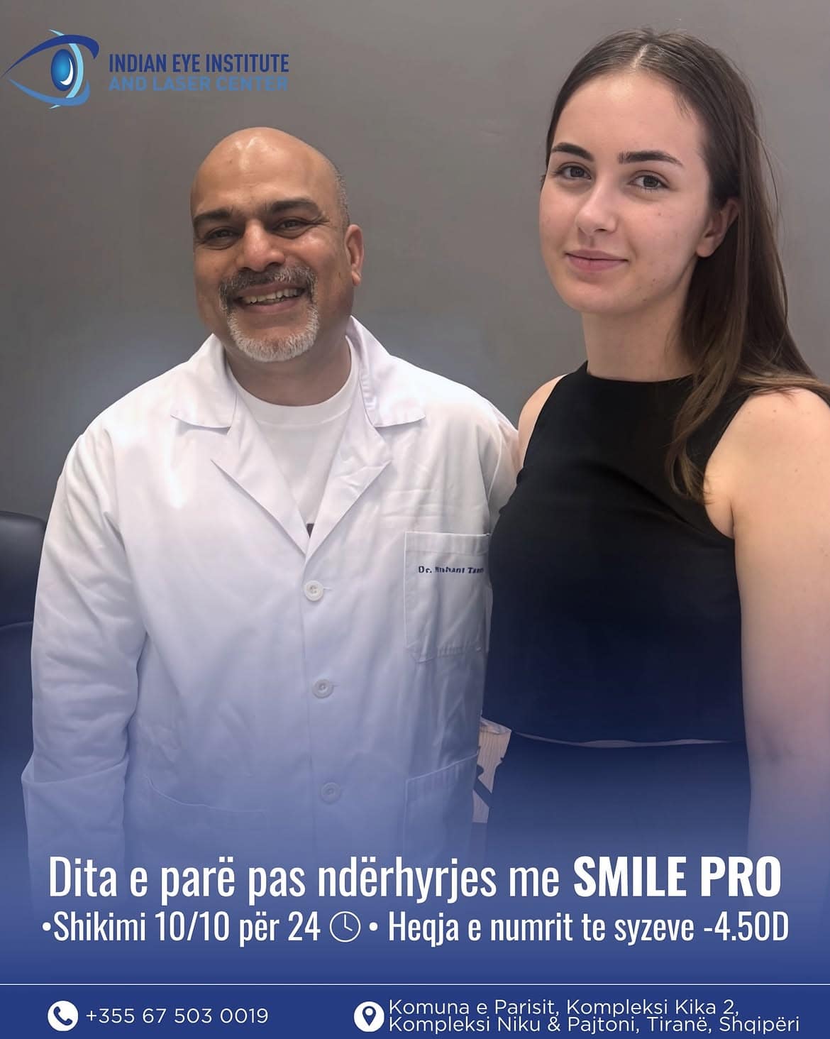
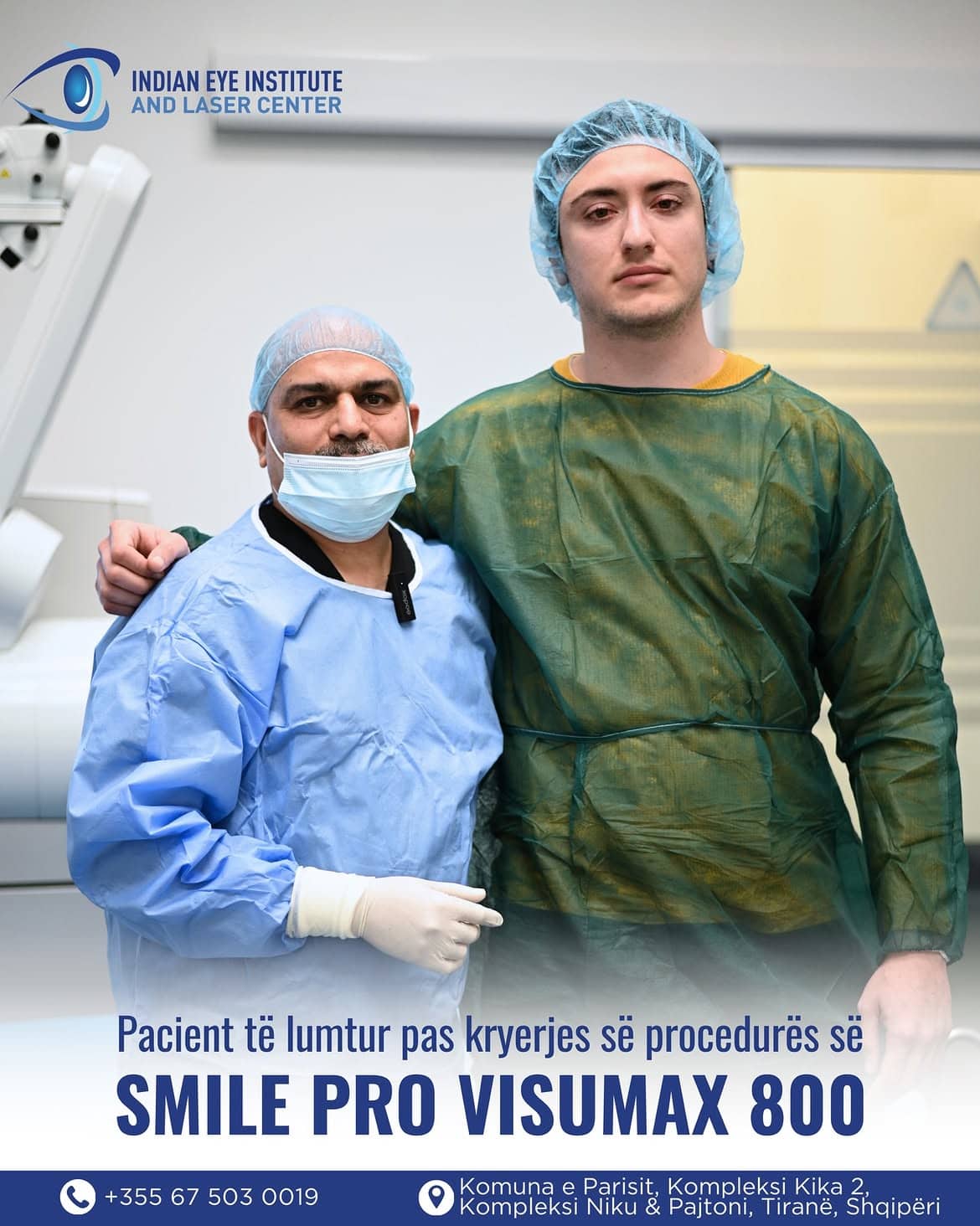
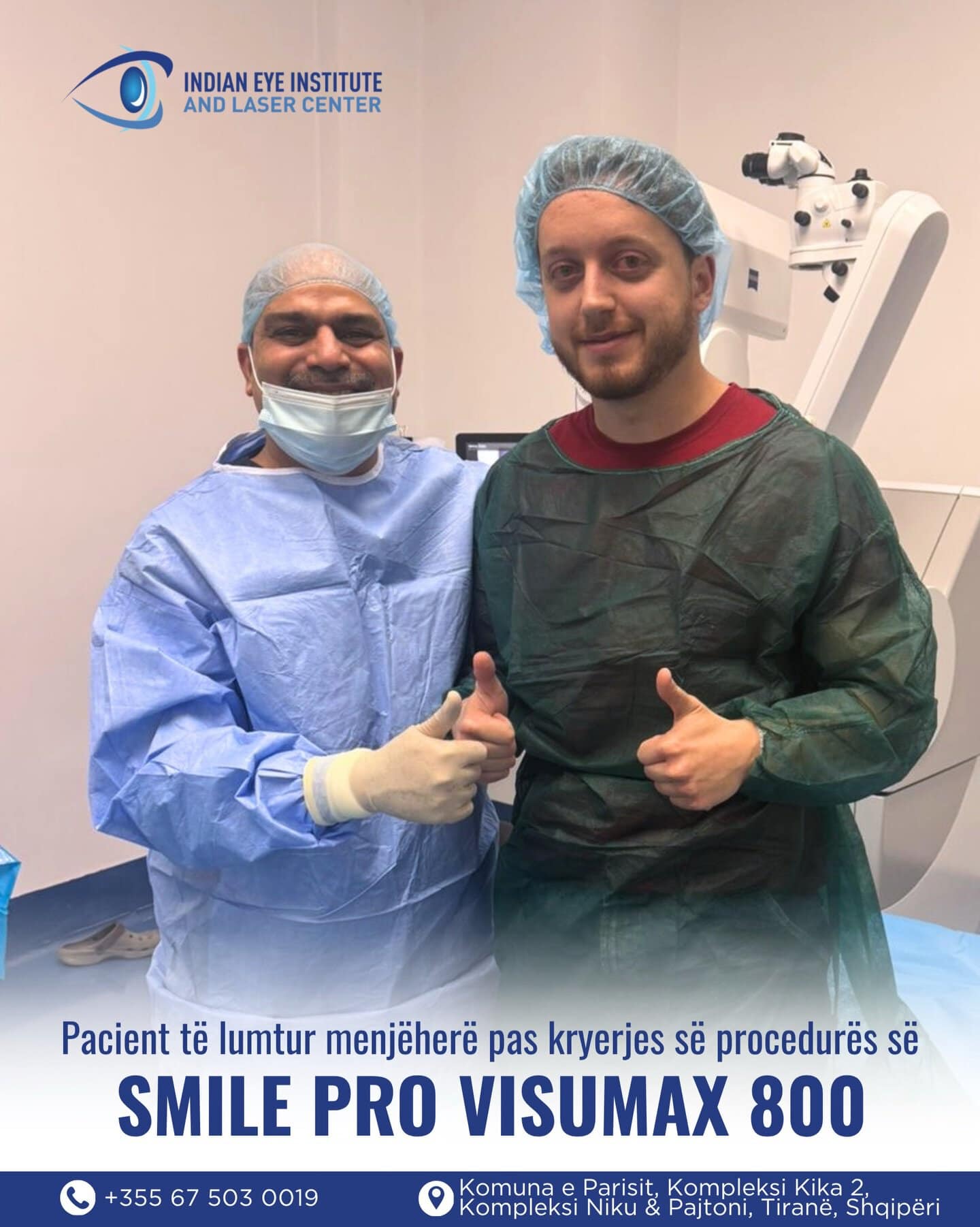
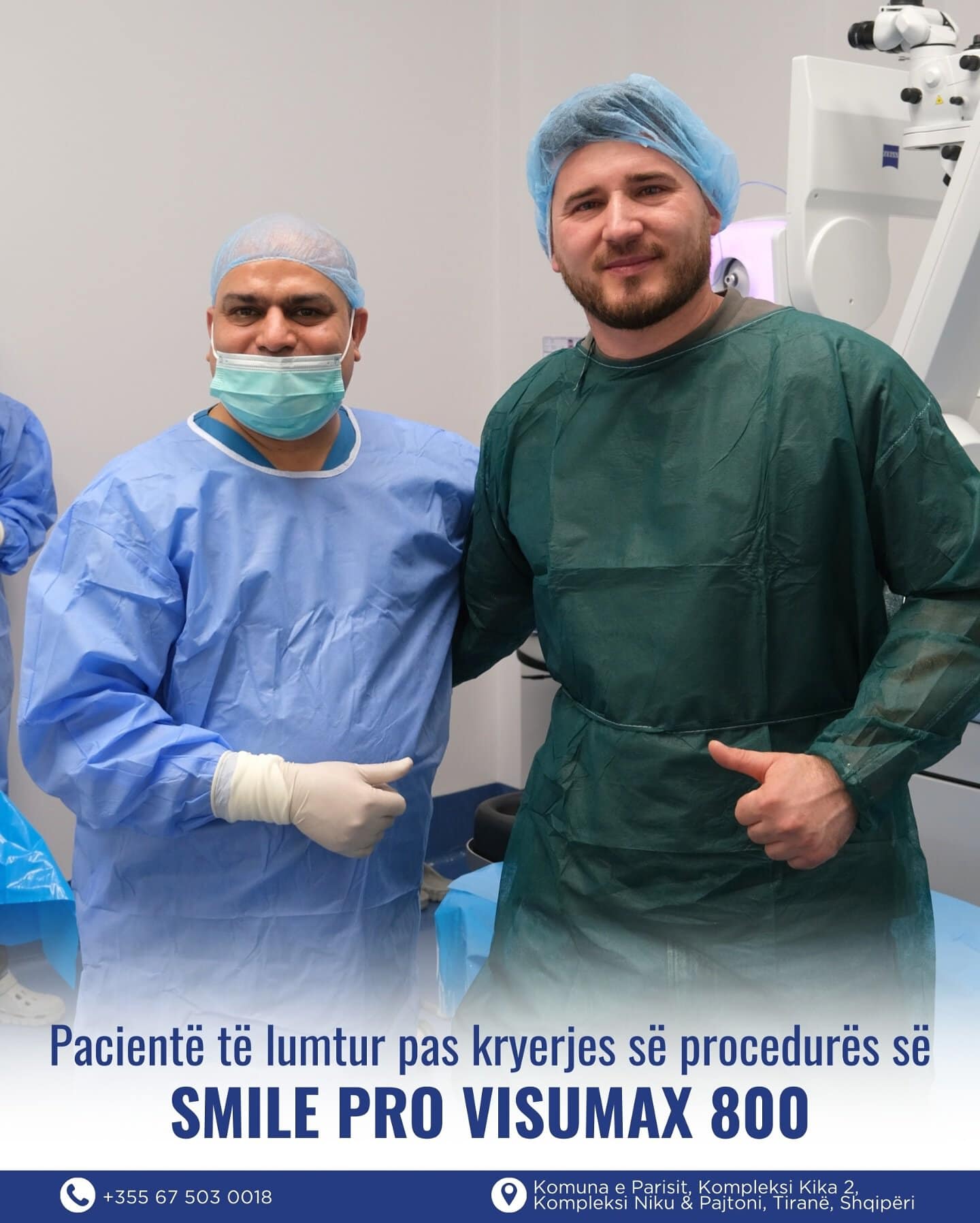
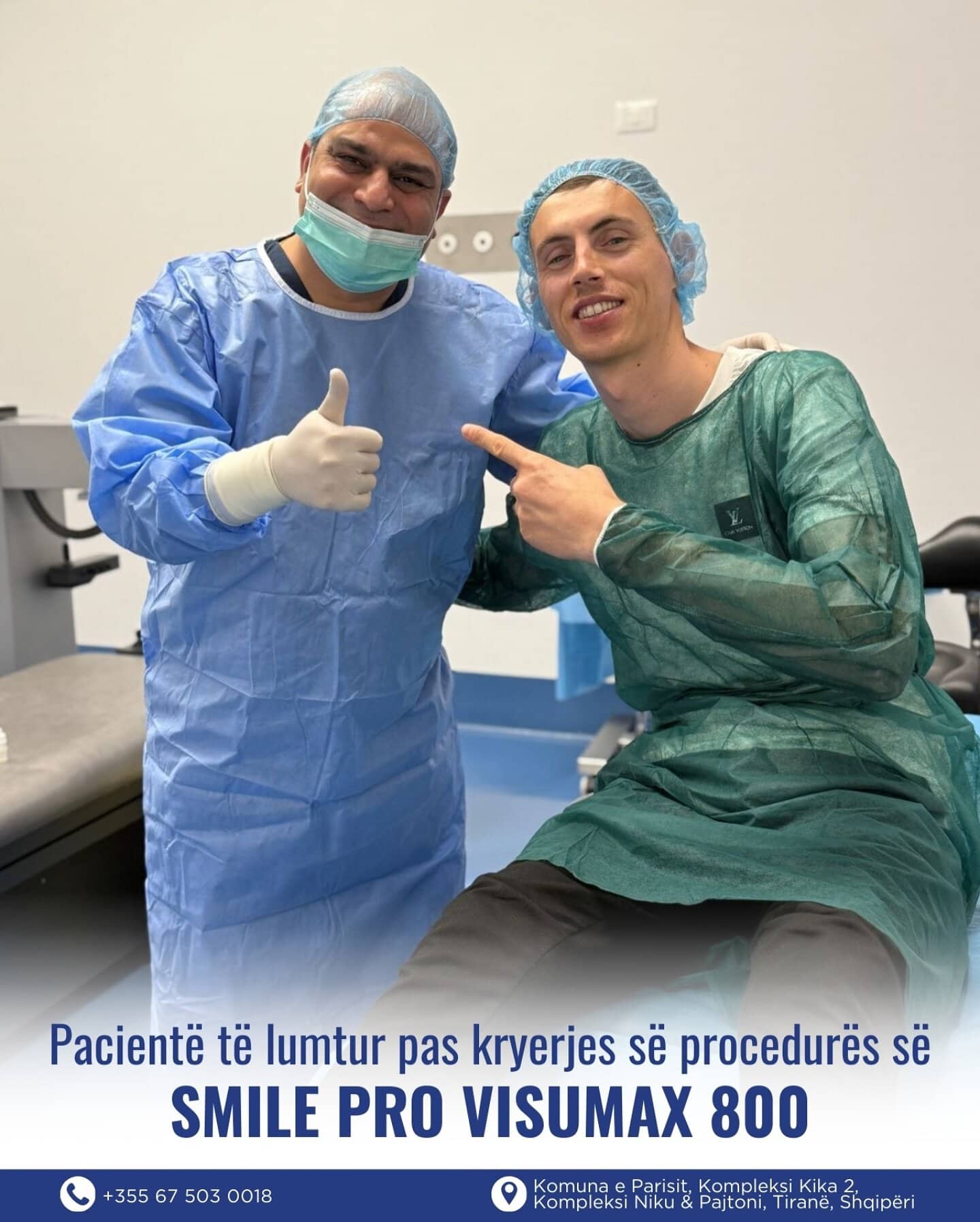
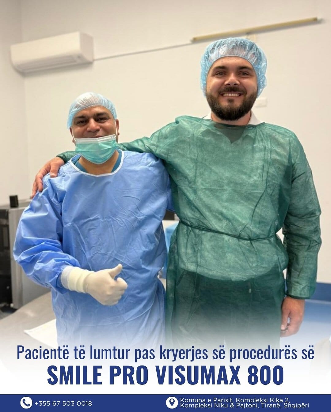
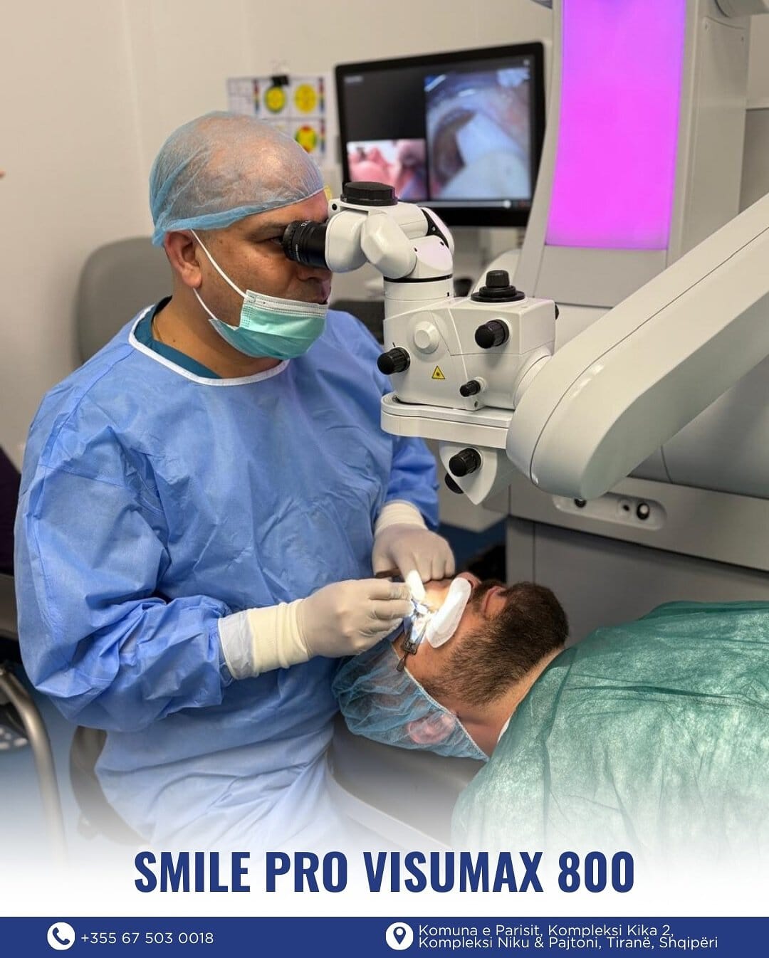
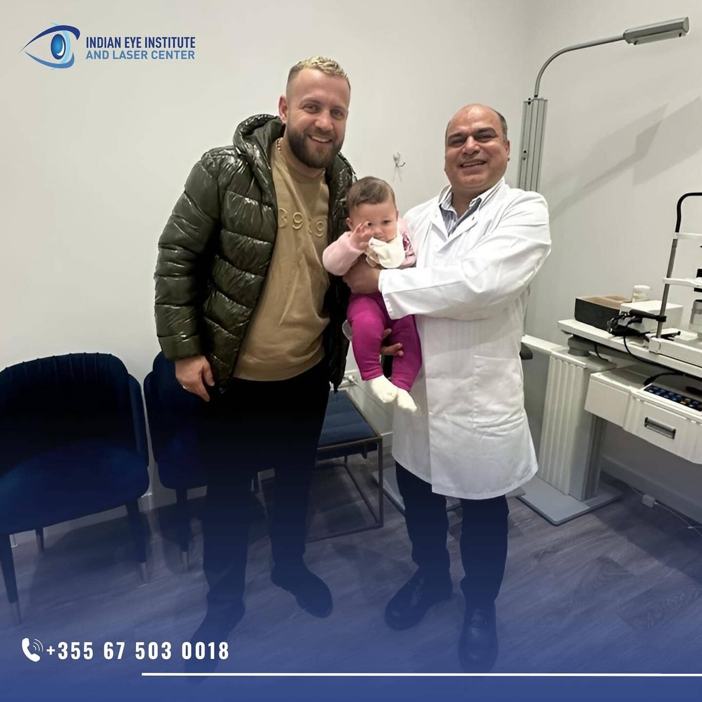
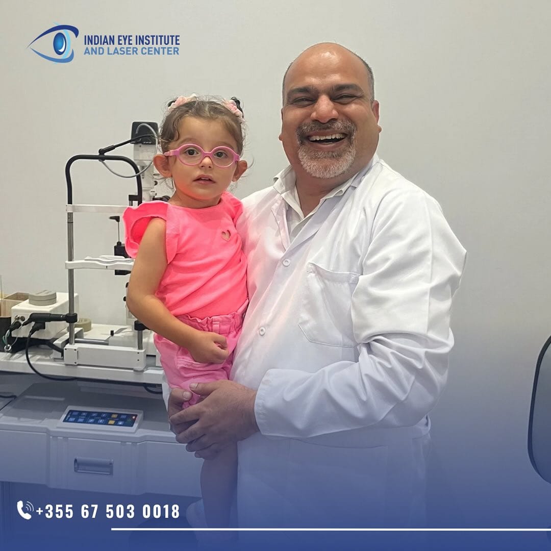
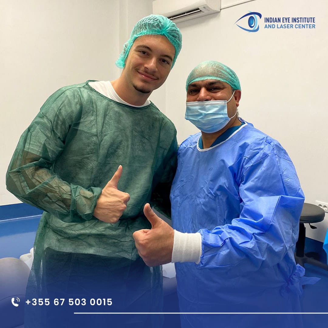
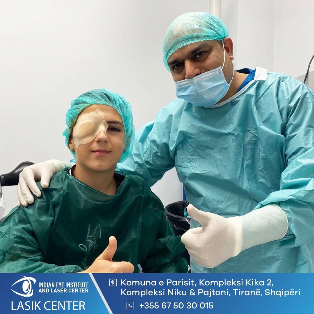
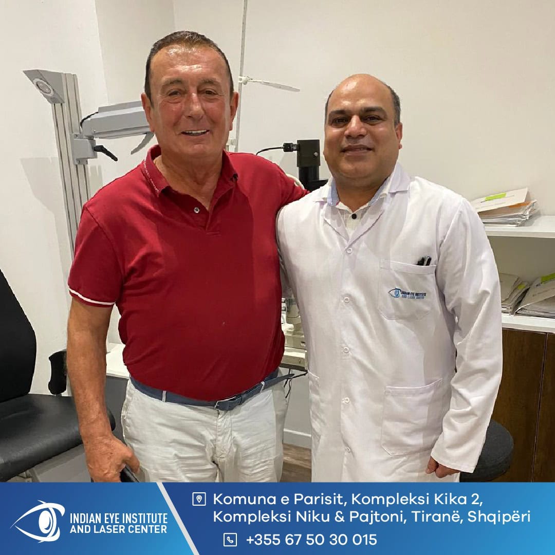
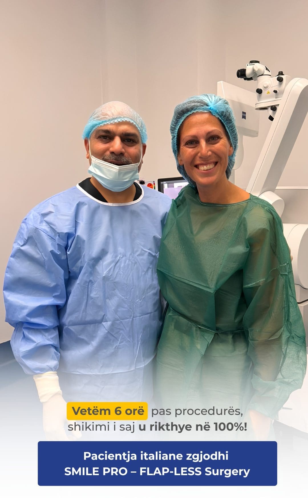
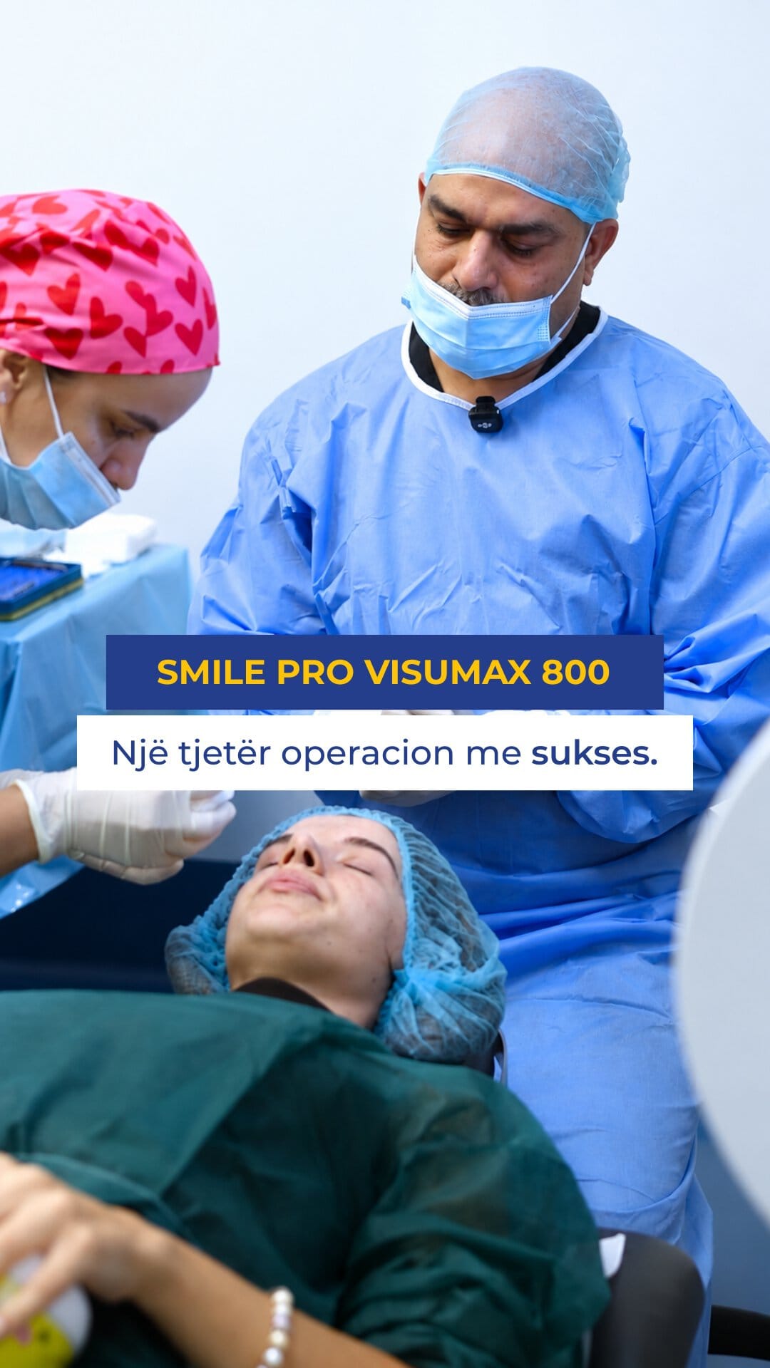
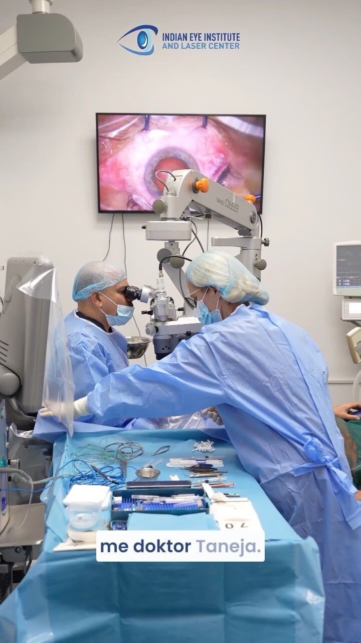
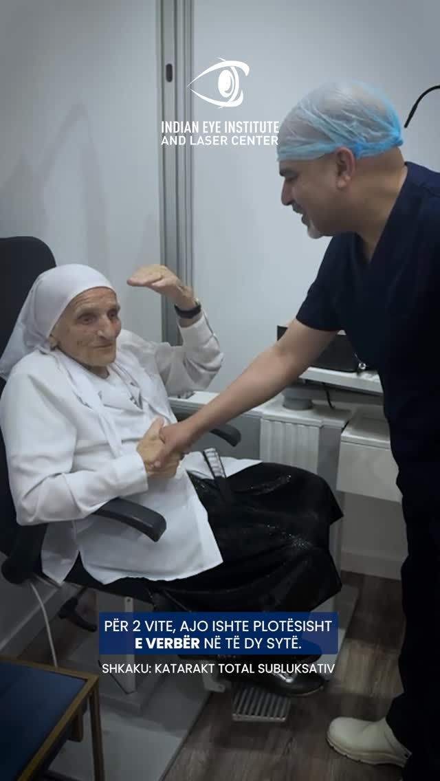
Moments with Our Patients












What Are the Symptoms of Keratoconus?
Symptoms vary depending on the stage and rate of progression of keratoconus. The most common signs include:
Blurred Vision
Visual Distortion
Frequent Changes in Prescription
Sensitivity to Light
Difficulty Seeing at Night
Double Vision or Ghost Images
Detection and Evaluation of Keratoconus
Keratoconus may be suspected when myopia and astigmatism change rapidly or when vision remains blurred despite correction with glasses. Examinations help confirm the diagnosis and monitor progression.
Vision and Refraction Assessment
The examination assesses visual acuity and changes in myopia or astigmatism. Frequent prescription changes may be a sign of keratoconus.
Corneal Topography
Topography creates a detailed map of the cornea’s shape and curvature. It helps identify steep and irregular areas, even in the early stages.
Corneal Measurement and Monitoring
Corneal thickness is measured and compared at follow-up appointments. Changes in shape, thickness, and steepness help determine whether keratoconus is progressing and whether corneal collagen cross-linking is needed.
Accurate diagnosis is particularly important in adolescents and young adults, as the condition may progress more rapidly in these age groups.
Can prevented Keratoconus?
Keratoconus cannot always be prevented because its exact cause is unknown. However, certain measures can help protect the cornea and detect progression early.
Avoid rubbing your eyes: Persistent, vigorous eye rubbing can further weaken the cornea and contribute to the progression of keratoconus.
Manage eye allergies: Allergy-related itching can lead to eye rubbing. Use only eye drops and treatments recommended by your doctor.
Have regular check-ups: Corneal topography and measurements help detect changes before vision deteriorates significantly.
Seek an eye examination if your prescription changes frequently: Rapid changes in myopia or astigmatism, particularly in young people, require a thorough corneal evaluation.
Benefits of Keratoconus Treatment
Treatment aims to improve the quality of vision and, when the disease is progressive, to strengthen the cornea and limit further deterioration. Outcomes depend on the stage of keratoconus and the treatment selected.
Early Diagnosis
Better Vision Correction
Corneal Strengthening
Stage-Specific Treatment
Management of Advanced Stages
Personalized Solutions for Keratoconus
Glasses and Specialized Contact Lenses
In the early stages, vision may be corrected with glasses. As corneal irregularity increases, rigid or hybrid contact lenses may be recommended to create a more regular optical surface.
- Prescription Glasses in the Early Stages
- Rigid Contact Lenses
- Hybrid Contact Lenses
- Regular prescription and contact lens fit checks
Corneal Collagen Cross-Linking
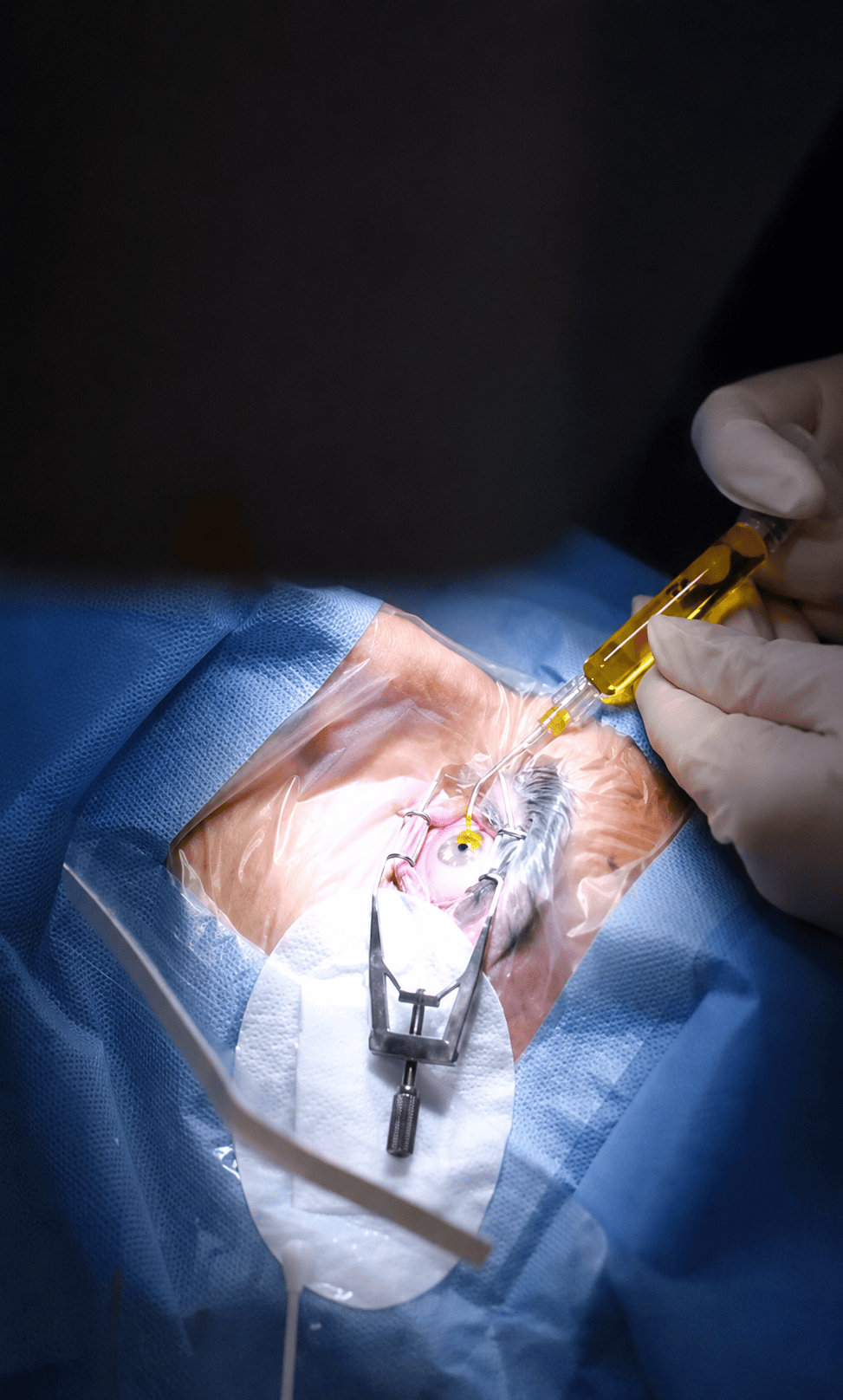
Corneal collagen cross-linking is used to strengthen the cornea and control the progression of keratoconus. During the procedure, riboflavin (vitamin B2) eye drops are applied and activated with ultraviolet light.
- Preparing the surface of the cornea
- Applying riboflavin eye drops
- Controlled exposure to ultraviolet light
- Placing a protective contact lens after treatment
Corneal Transplantation
Corneal transplantation is considered only in advanced cases, when glasses and contact lenses no longer provide adequate vision or when the cornea has become severely thinned or damaged.
- Partial-thickness corneal transplant
- Full-thickness corneal transplant
- Replacing damaged tissue with donor corneal tissue
- Ongoing follow-up during recovery
What to Expect
01. Comprehensive Corneal Examination
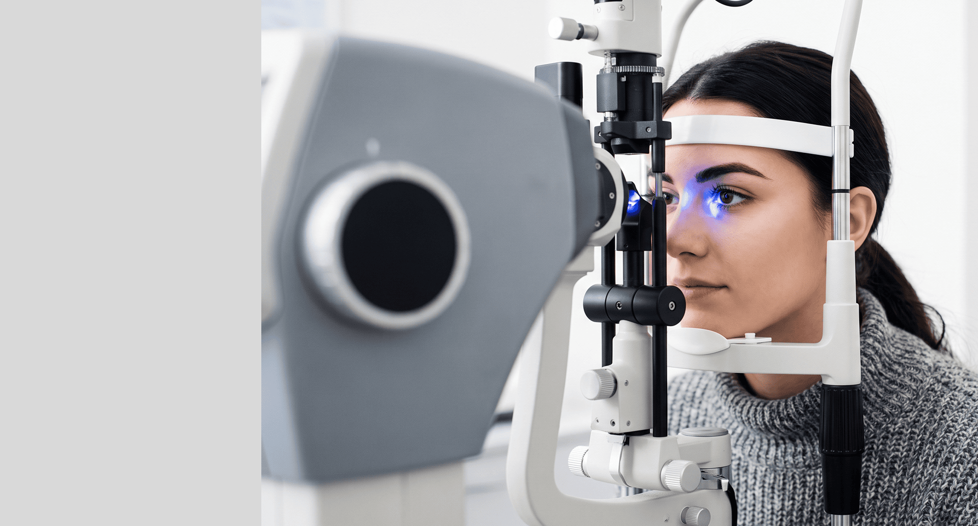
During the examination, visual acuity, myopia, and astigmatism are assessed. The cornea is examined with a slit-lamp biomicroscope to identify thinning, changes in shape, or signs of damage.
Corneal topography creates a detailed map of the corneal surface. Measuring corneal thickness helps determine the stage of the condition and assess suitability for corneal collagen cross-linking.
The results are compared during regular follow-up visits to determine whether the keratoconus is stable or progressing.
02. Procedure
Corneal collagen cross-linking is performed as an outpatient procedure under local anesthesia using anesthetic eye drops. The surface layer of the cornea is carefully removed before treatment.
Riboflavin drops are applied to the cornea. Controlled ultraviolet light is then used to activate the riboflavin and help create stronger bonds between the existing collagen fibers.
The procedure takes about one hour. At the end, a protective contact lens is placed over the eye and remains in place until the surface of the cornea has healed.
03. Recovery and Follow-Up
During the first few days, patients may experience pain, tearing, sensitivity to light, a scratchy sensation, and blurred vision. The protective lens is usually worn for up to three days or until the surface of the cornea has healed.
Antibiotic eye drops are used while the surface of the eye heals, while steroid eye drops may be used for several weeks according to the schedule provided by the clinic.
Many patients can return to work after about one week, although vision may fluctuate for several weeks or months. Patients should avoid rubbing the eye, dust, swimming, and strenuous activities for the recommended period.
Worsening pain, discharge, severe redness, or a sudden decrease in vision requires immediate contact with the clinic.


Indian Eye Clinic Tirana: 15+ Years of Trust


Retina | Cataract | Refractive Surgery


The right care for a lifetime with clear vision
Every successful treatment begins with an accurate diagnosis and a personalized care plan. Discover the real stories of patients who trust Indian Eye Institute and Laser Center.





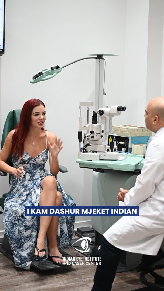
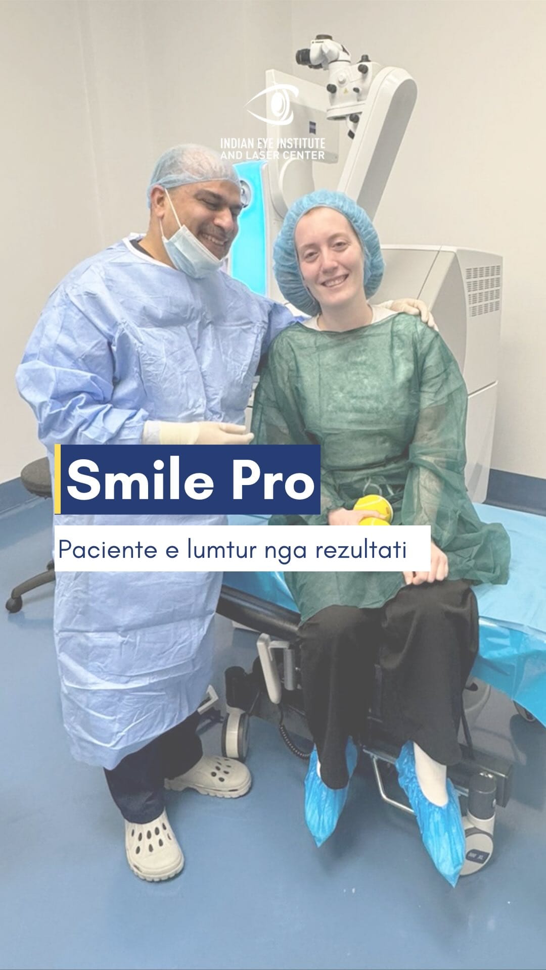
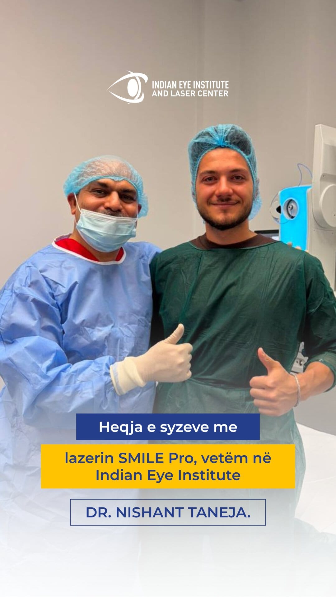


I have advanced glaucoma in both eyes and have undergone glaucoma and cataract surgery with Dr. Taneja. I have been receiving regular follow-up care from Dr. Taneja for the past 11 years.
I am very happy and satisfied with my experiences at Dr. Taneja’s clinic. The staff are helpful, knowledgeable, competent, and very pleasant. Dr. Taneja has worked diligently to preserve my vision, and I am extremely pleased with the care I have received. He deserves recognition for how good he is at what he does. I would recommend him 100% to anyone with serious eye problems.
I have suffered from a long-term problem with floaters in both eyes. Dr. Nishant Taneja is truly an expert in his field who allows patients to consider nonsurgical options as the first course of treatment. He is professional and courteous, treats patients with respect, and communicates clearly in terms that ordinary people can understand. I believe he is honest and straightforward, and he is taking excellent care of me.
I have been a patient of Dr. Nishant Taneja since 2010, when I was diagnosed with wet macular degeneration. He has treated me with Avastin, Eylea, and now brolucizumab, and I can still read large print.
I owe my sight to his treatments. He has taken excellent care of my eyes. The staff are absolutely outstanding in their courtesy and professionalism. I am 93 years old and can still read clearly. Thank you!
When I first came here, I thought I would go blind in my left eye. I was diagnosed with a vitreous hemorrhage caused by detachment of the vitreous gel. After the laser treatment, my vision returned to what it had been before.
I have received excellent medical care here. The entire staff have been very kind and have made everything much easier for me. I am very happy.
Many thanks to the entire staff. The whole process of my cataract surgery and the care I received were the best I have ever experienced at any medical practice. The staff and technology were first-rate. Dr. Taneja is simply phenomenal. Thank you again.
I underwent surgery for a retinal detachment last week. I experienced no pain during or after the operation. I slept well the following night without needing any pain medication. If anyone has questions about retinal surgery, please feel free to call me.
I am impressed by the high standard of patient care I received from Dr. Taneja at the Indian Eye Institute. I hold him in the highest regard.
I have had diabetes for over 30 years. Over the past 5 years, my vision gradually deteriorated. I had been receiving treatment from my ophthalmologist for the previous 4 years. The treatment involved injections, but the situation seemed to be getting worse. I could barely see well enough to drive, could not read anything on my phone, and began having difficulty recognizing people from a distance. I therefore decided to visit Dr. Nishant Taneja on the recommendation of my eye doctor. He was prompt, efficient, and professional. Within minutes, he had mapped out a treatment plan for both of my eyes and advised me to follow it. I was truly impressed by his energy, optimism, and confidence.
After several injections and laser treatments, he said that I needed a vitrectomy in my left eye. It went perfectly, without any complications. My vision in that eye was restored to 20/20. I felt as though I were opening my eyes for the first time.
After I recovered from that procedure, Dr. Taneja said that my right eye also needed surgery. So, without hesitation, I had that eye treated as well. I now have 20/20 vision in both eyes, and everything is wonderful.
I can now see my two young grandchildren clearly. This has improved my quality of life tremendously. Without a doubt, I am probably the happiest patient Dr. Taneja has ever had!
I am delighted to express my appreciation for Dr. Taneja and his exceptional staff.
If you are reading this review while deciding on a retina specialist, you have definitely come to the right place! An excellent doctor and staff. I am more than completely satisfied!
I believe Dr. Taneja is an exceptionally talented surgeon.
I began wearing glasses and contact lenses at the age of 12. I relied on them to see and read. During the spring, pollen irritated my eyes, and I could no longer wear my contact lenses.
After the procedures performed by Dr. Taneja, I went from being unable to see to having clear vision without contact lenses or glasses.
I would recommend Dr. Nishant Taneja for any procedure he performs. He is highly meticulous, compassionate, and understanding.
He worked a miracle for me. I thank God for his talent.
Patient Education Videos
View All
The SMILE Pro Procedure
Chemical Eye Injuries
Diabetic Retinopathy
Signs That Should Not Be Ignored
Examination and Treatment
Download the Guides Free for Patients


Frequently Asked Questions
The exact cause is unknown, but keratoconus may have a genetic component. People with close relatives who have keratoconus may be advised to undergo a corneal examination, particularly if they have astigmatism that changes frequently.
Standard LASIK surgery is generally not recommended for patients with keratoconus because removing tissue may further weaken the cornea. Any vision correction option should be evaluated by a specialist after corneal topography and corneal thickness measurements.
Anesthetic eye drops are used during the procedure, and the patient usually does not feel any pain. Once the anesthesia wears off, pain, a scratchy sensation, and sensitivity to light may occur for several days.
Corneal collagen cross-linking is primarily intended to stop the progression of keratoconus and does not guarantee that corrective lenses will no longer be needed. Many patients continue to use glasses or contact lenses to achieve the best possible vision.
Take the First Step Toward Better Vision.
Fill out the form below and our team will contact you
to schedule a consultation and answer your questions.